Arthritis and Other Abnormalities of the Talonavicular Joint
Anatomic Considerations
The talonavicular joint, which together with the calcaneonavicular joint make up Chopart’s joint, which represents the posterior border of the midfoot (Fig. 3.1). The talonavicular joint comprises articulation of the talus and the navicular bone (Fig. 3.2). The articular surfaces are covered with hyaline cartilage, which are susceptible to arthritis. Most of the strength of the talonavicular joint is provided by the talonavicular and plantar calcaneonavicular ligaments (Fig. 3.3). The joint capsule is lined with a synovial membrane that attaches to the articular cartilage and may give rise to bursae. In addition to arthritis, the tibiofibular joint is susceptible to the development of tendinitis, bursitis, and disruption of the ligaments, cartilage, and overlying tendons.
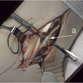
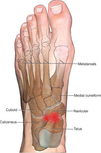 FIGURE 3.1 The articulations of the talonavicular joint. |
Clinical Correlates
The talonavicular joint, which together with the calcaneonavicular joint make up Chopart’s (also known as the transmetatarsal joint) joint, which represents the posterior border of the midfoot (Fig. 3.1). The talonavicular joint comprises the articulation of the talus and the navicular bone. The joint’s articular cartilage is susceptible to damage, which left untreated will result in arthritis with its associated pain and functional disability. Osteoarthritis of the joint is the most common form of arthritis that results in talonavicular joint pain and functional disability, with rheumatoid arthritis and posttraumatic arthritis also causing arthritis of the talonavicular joint (Fig. 3.4). The joint is susceptible to strains and sprains due to the stresses placed on the midfoot especially when engaging in high-impact activities such as running on hard surfaces with inadequate footwear (Fig. 3.5). Less common causes of arthritis-induced talonavicular joint pain include the collagen vascular diseases, infection, villonodular synovitis, and Lyme disease. Acute infectious arthritis of the talonavicular joint is best treated with early diagnosis, with culture and sensitivity of the synovial fluid and prompt initiation of antibiotic therapy. The collagen vascular diseases generally manifest as a polyarthropathy rather than a monoarthropathy limited to the talonavicular joint, although talonavicular pain secondary to the collagen vascular diseases responds exceedingly well to ultrasound-guided intra-articular injection of the talonavicular joint.
Patients with talonavicular joint pain secondary to arthritis and collagen vascular disease related joint pain complain of pain that is localized to the anterior ankle and midfoot. Activity, especially involving inversion and eversion of the talonavicular joint makes the pain worse, with rest and heat providing some relief. The pain is constant and characterized as aching. Sleep disturbance is common with awakening when the patient rolls over onto the affected talonavicular joint. Some patients complain of a grating, catching, or popping sensation with
range of motion of the joint, and crepitus may be appreciated on physical examination.
range of motion of the joint, and crepitus may be appreciated on physical examination.
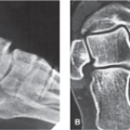
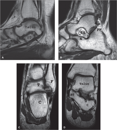 FIGURE 3.2 T1-weighted MR images demonstrating hindfoot and midfoot anatomy. A: Sagittal image at the flexor digitorum longus level (FDL). B: Sagittal image at talonavicular level demonstrating the posterior talocalcaneal facet (PF), the tarsal sinus (TS), and the anterior calcaneal process (A). C: Coronal image at the level of the lateral ankle mortise. D: Coronal image at the level of the posterior medial ankle mortise. N, navicular; ST, sustentaculum tali; C, calcaneus; T, talus; F, fibula; PTFL, posterior talofibular ligament; TS, tarsal sinus; ST, sustentaculum tali; DL, deltoid ligament. (From Berquist TH. Anatomy, normal variants, and basic biomechanics. In: Imaging of the Foot and Ankle. 3rd ed. Philadelphia, PA: Lippincott Williams & Wilkins; 2011:1–46.) |
Functional disability often accompanies the pain associated with the many pathologic conditions of the talonavicular joint. Patients will often notice increasing difficulty in performing their activities of daily living and tasks that require walking, climbing stairs, and walking on uneven surfaces are particularly problematic. If the pathologic process responsible for the patient’s pain symptomatology is not adequately treated, the patient’s functional disability may worsen and muscle wasting may occur.
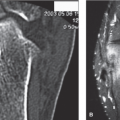
Plain radiographs are indicated in all patients who present with talonavicular pain as not only intrinsic talonavicular disease as well as other regional pathology may be perceived as talonavicular pain by the patient (Figs. 3.4 and 3.6). Based on the patient’s clinical presentation, additional testing may be
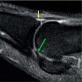
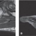
indicated, including complete blood cell count, sedimentation rate, and antinuclear antibody testing. MRI, CT, or ultrasound of the talonavicular joint is indicated if avascular necrosis or meniscal tear is suspected or if the diagnosis is unclear (Fig. 3.7).
indicated, including complete blood cell count, sedimentation rate, and antinuclear antibody testing. MRI, CT, or ultrasound of the talonavicular joint is indicated if avascular necrosis or meniscal tear is suspected or if the diagnosis is unclear (Fig. 3.7).
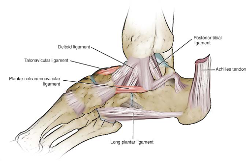 FIGURE 3.3 The articulations of the talonavicular joint.
Related posts:Stay updated, free articles. Join our Telegram channel
Full access? Get Clinical Tree
 Get Clinical Tree app for offline access

|