Key Points
- •
Point-of-care ultrasound is defined as a goal-directed, bedside ultrasound examination performed by a healthcare provider to answer a specific diagnostic question or to guide performance of an invasive procedure.
- •
Diagnostic ultrasound was first developed and used in medicine during the 1940s, but point-of-care ultrasound has been integrated into diverse areas of clinical practice since the early 1990s.
- •
Important considerations when using point-of-care ultrasound include provider training and skill level, patient characteristics, and ultrasound equipment features.
Background
Point-of-care ultrasound has revolutionized the practice of medicine, influencing how care is provided in nearly every medical and surgical specialty. For more than a century, clinicians had been limited to primitive bedside tools, such as the reflex hammer (c. 1888) and stethoscope (c. 1816), but with bedside ultrasound, providers are equipped with a tool that allows them to actually see what they can only infer through palpation or auscultation. The technologic miniaturization of ultrasound devices has outpaced integration of these devices into clinical practice. Many professional societies and national organizations have recognized the potent impact of point-of-care ultrasound and have endorsed its routine use in clinical practice. In 2001 the American Medical Association stated, “Ultrasound has diverse applications and is used by a wide range of physicians and disciplines. Ultrasound imaging is within the scope of practice of appropriately trained physicians.” Thus, it has been well recognized for over a decade that providers from diverse specialties can and should be trained to use ultrasound within their scope of practice. This chapter reviews the major milestones in the history of medical ultrasound with a focus on important considerations of point-of-care ultrasound.
History
Acoustic properties of sound were well described by ancient Greek and Roman civilizations. In the twentieth century, the sinking of the Titanic followed by the start of World War I served as catalysts for the development of sonar, or sound navigation and ranging, which was the first real-world application of the principles of sound.
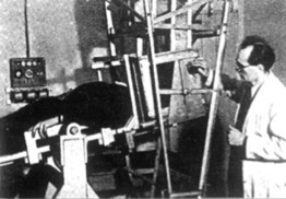
Although several physicians were simultaneously competing for recognition as the first to use ultrasound in medicine, Karl Theodore Dussik, an Austrian psychiatrist and neurologist, is credited as being the first physician to use ultrasound in medical diagnostics when he attempted to visualize cerebral ventricles and brain tumors using a primitive ultrasound device in 1942 ( Figure 1.1 ).
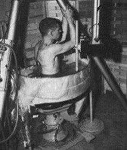
During the 1940s and 1950s, many pioneers advanced the field of medical ultrasound. John Julian Wild described various clinical applications of ultrasound, including the difference in appearance of normal and cancerous tissues. Douglass Howry and Joseph Holmes focused on ultrasound equipment technology. They built immersion-tank ultrasound systems, including the “somascope” in 1954 ( Figure 1.2 ), and they published the first two-dimensional ultrasound images. Ian Donald contributed significant amounts of research in obstetric and gynecologic ultrasound. Inge Edler and Carl Hellmuth Hertz investigated cardiac ultrasound and established the field of echocardiography in the early 1950s. Shigeo Satomura, a Japanese physician isolated from the pioneers in the United States and Europe, is credited as being the first physician to use Doppler ultrasound in his studies of cardiac valve motion.
Advancements in ultrasound technology accelerated the field in the 1960s and 1970s. Early ultrasound machines used open-shutter photography to capture screen images. Multiple still images of moving structures were captured, sequentially displayed, and interpreted by trying to imagine the structures in motion. In 1965, Siemens released the Vidoson, the first real-time ultrasound scanner, which was able to display 15 images per second. The Vidoson was quickly incorporated into obstetric care over the next decade and became a standard component of assessing pregnant women. Sector scanning became possible with development of phased-array transducers in the early 1970s, giving rise to echocardiography as an independent field.
Ultrasound technology continued to advance during the 1970s and 1980s with the development of more sophisticated transducers along with refinements in image quality. Following the “early adopters” of ultrasound, namely, radiology, cardiology, and obstetrics/gynecology, ultrasound began to be used in emergency care, a role that marked the beginning of the era of point-of-care ultrasound use by healthcare providers from diverse medical specialties. Life-threatening conditions could be assessed rapidly at the bedside with portable ultrasound. Frontline physicians, mostly surgeons and emergency medicine physicians, started assessing trauma patients with ultrasound in the 1970s, and the term FAST exam, or Focused Assessment with Sonography in Trauma, was coined in the early 1990s. The FAST exam was incorporated into Advanced Trauma Life Support (ATLS) guidelines in the late 1990s. From its early description in the 1970s in Europe to its incorporation into ATLS guidelines in the 1990s in the United States, the FAST exam established a precedent for development of point-of-care ultrasound applications and incorporation of these applications into routine clinical practice.
Since the 1990s, point-of-care ultrasound has become a part of nearly every specialty’s practice. In addition to development of specific point-of-care ultrasound applications in the 1990s, such as the FAST exam, general medical ultrasound applications, broadly applicable to many specialties, started to emerge. In the mid-1980s, ultrasound artifacts of the lung and pleura—an organ long felt to have little utility in ultrasound diagnostics—began to be described. The identification and codification of these artifacts with discrete forms of lung pathology was developed by Daniel Lichtenstein, a French critical care physician, which gave rise to the field of lung ultrasonography. Even though lung ultrasound was first used by intensivists to evaluate critically ill patients, lung ultrasound is broadly applicable to evaluate any patient with pulmonary symptoms, is more accurate than chest x-ray, and can be used by any healthcare provider, regardless of specialty. Another broadly applicable use of ultrasound that evolved was in the guidance of invasive bedside procedures. Multiple studies since the 1990s have demonstrated reduced mechanical complications when ultrasound is used to guide common procedures, in particular central venous catheterization. Current guidelines recommend that all providers, regardless of specialty, use ultrasound guidance when placing internal jugular central venous catheters.
Ultrasound technology was well advanced by the 2000s, which saw the development of three-dimensional ultrasound for select diagnostic applications; however, two-dimensional ultrasound has remained the standard for the majority of indications. The more important change during the 2000s was continued reduction in the size and price of ultrasound machines. The increased portability and affordability of ultrasound devices led to an exponential increase in use of ultrasound by all providers. Subsequently, many professional societies published guidelines on use of point-of-care ultrasound, including the American Institute of Ultrasound in Medicine (AIUM), American College of Emergency Physicians (ACEP), American College of Chest Physicians (ACCP), and American Society of Echocardiography (ASE). Furthermore, consensus guidelines between imaging and specialty societies have been established, such as the guidelines on obstetrical ultrasound collaboratively developed by the American College of Radiology (ACR), American College of Obstetricians and Gynecologists (ACOG), American Institute of Ultrasound in Medicine (AIUM), and Society of Radiologists in Ultrasound (SRU) and the ACEP–ASE statement on focused cardiac ultrasound in the emergent setting. Specialty-specific guidelines also emerged, such as the American Association of Clinical Endocrinologists guidelines on thyroid ultrasound that defined a pathway for endocrinologists to earn a certificate of competency in thyroid and neck ultrasound.
Medical educators recognized the importance of a basic understanding of ultrasound in the early 2000s and started to explore how to incorporate ultrasound training into curricula for medical students, residents, and fellows. The Accreditation Council for Graduate Medical Education (ACGME) has started to mandate certain residency and fellowship programs in the United States include basic ultrasound education; for example, critical care ultrasound and ultrasound-guided thoracentesis and central venous catheterization are now required components of pulmonary/critical care fellowship training. Several medical schools worldwide have started to expose their students to the principles and practice of ultrasound, most often in conjunction with anatomy and physical examination courses. The coming generation of physicians will thus be more adept at point-of-care ultrasound applications and will consider use of bedside ultrasound to be routine in most clinical encounters. While past generations’ contributions established the utility of ultrasound as a valuable bedside tool in diagnostics and procedural guidance, the next generation will advance the field by studying how point-of-care ultrasound can be best incorporated into patient care algorithms and its impact on healthcare outcomes, cost-effectiveness, and patient satisfaction.
Related posts:


Stay updated, free articles. Join our Telegram channel

Full access? Get Clinical Tree


