Film Interpretation and Report Writing

Chapter 5


Film Interpretation and Report Writing

Dennis M. Marchiori

Matthew Richardson

FOCUS ON RADIOGRAPHS

COMMON USES OF RADIOGRAPHS

CRITERIA FOR ORDERING RADIOGRAPHS

IMAGE INTERPRETATION

REPORT WRITING

SUMMARY

SAMPLE RADIOLOGY REPORTS

Focus on Radiographs

Since its inception, diagnostic imaging has played a fundamental role in patient evaluation. Diagnostic imaging began more than 100 years ago with the advent of plain film radiography, and has progressed to advanced modalities, such as digital radiography, magnetic resonance imaging (MRI), computed tomography (CT), and positron emission tomography (PET). Diagnostic imaging remains an essential tool to recognize, define, identify, and exclude many of the common and not-so-common pathologies encountered in a health care setting.

For most clinical assessments, the historically dominant plain film radiography continues to be the first step, with more sophisticated specialized imaging systems applied as follow-ups to negative, equivocal, or ambiguous results from the plain film study, or for clinical questions for which plain films are known to be insensitive. Plain film radiology is widely available, relatively inexpensive, and rapidly obtained, suggesting it will remain the most common imaging method for the near future.

Plain film radiology represents a common denominator between many health professions. Dentists, podiatrists, chiropractors, medical physicians, and many practitioners in the allied health professions routinely rely on information obtained from plain film radiographs to manage their patients. With this in mind, this chapter focuses on the interpretation and reporting of plain film radiographs. However, with little modification, the concepts presented here and targeted to plain film radiology equally relate to other specialized imaging modalities. Consequently, this chapter’s goal is to provide the reader with an understanding of issues related to when radiographs should be taken, methods to successfully search radiographs for abnormal findings, the ability to categorize these findings by appearance and location into common patterns, and steps to summarize and report on these findings successfully.

Common Uses of Radiographs

In 2009, an estimated $2.5 trillion was spent on personal health care in the United States.1 Health-care expenditure was 17.6% of the gross domestic product, representing a per capita expenditure of $8,086.1 Imaging is a growing portion of these costs. In 1990, an estimated 3.5% of health-care expenditures were for radiologic services.2,3 This percentage had grown to 9.1% in 2005, ranking it as the third largest health-care expenditure category after hospital care and physician services.4

Diagnostic imaging is common to clinical practice. Historically, more than 80% of chiropractors use radiographs as part of their clinical protocol and have the necessary equipment to produce radiographs in their offices.5 Although national data are not available, a survey of Minnesota medical physicians found that approximately 87% had onsite radiology equipment.6

Developing film interpretation skills is of obvious interest to radiologists, but developing these skills is also important to nonradiologist medical and chiropractic physicians, and other health-care providers who often take and interpret radiographs as part of patient evaluation and management. For instance, most chiropractors do not regularly consult with radiologists to assist their interpretations. In fact, fewer than 20% of hospitals have full-time onsite coverage by a board-certified radiologist.7 During these off hours, the initial interpretation and related decisions are often done by nonradiologist clinicians, most to be read by radiologists later. Moreover, of the radiologic services done in a private medical practice setting, 57%2,3 to 70%8,9 are performed and interpreted by nonradiologists.

Questions arise related to the appropriateness of the training of nonradiologists to interpret imaging and under what circumstance it is best to consult with a radiologist. The American College of Radiology (ACR) recommends that radiographs be interpreted by certified radiologists or physicians who have documented training in an approved residency, including radiographic training on all body areas. This indicates the basic need for formal training, but does not limit interpretations to radiologists.10 Literature within the chiropractic profession advocates for greater use of chiropractors who are certified with advanced training in radiology (Diplomates of the American Chiropractic Board of Radiology [DACBR]) as a method of limiting liability and enhancing accuracy of image interpretation.11

Taylor12 found that the degree of training positively influences the ability of medical and chiropractic clinicians and students to correctly identify selected bone and joint pathology. As one might expect, the concordance between the radiographic interpretation of radiologists and nonradiologists is best for extremity bone radiographs (approximately 95%) and lower for more complex studies, such as chest radiographs (generally ranging from 40% to 90%).*

Criteria for Ordering Radiographs

The most effective application of diagnostic imaging for common clinical presentations is widely debated. The multifaceted and unique clinical presentations of most patients make the formation of common criteria for ordering diagnostic imaging challenging. Everyone agrees that all radiographic examinations should follow clear historical and clinical indications because of examination costs and the potentially hazardous effect of ionizing radiation.13 Unfortunately, there is no general agreement on exactly what these historical and clinical indications should be to satisfy the competing needs of gaining information while limiting cost and radiation exposure.

To date, diverse opinion exists about what constitutes accepted clinical criteria for ordering radiographs for patients with musculoskeletal complaints. Although the literature contains many attempts to develop criteria for ordering radiographs for patients with complaints of the spine, no system has been generally accepted. As a matter of observation, the use rates of plain film radiographs vary widely. Developing and embedding guidelines seem easier tasks for a narrow-scope presentation for something such as ankle14,15 or knee trauma,16,17 but are less successful for case presentations of increasing complexity and ambiguity (e.g., back pain) and applied management (e.g., pharmaceuticals versus manual adjustments or manipulation of the spine).

Opinions vary widely about the use of radiographs in the evaluation of patients experiencing back pain. Multiple questions impact the issue. Should radiographs be taken for patients who are experiencing acute but not chronic back pain? What are the appropriate film-ordering criteria that maximize clinical information yet minimize patient cost and radiation exposure?

Despite the fact that these topics have garnered considerable attention over the past decade, evidence-based guidelines for the use of plain film radiology (or CT and MRI) are not widely used in the clinical setting. In 1987 the Quebec Task Force, and later the United States’ Agency for Health Care Policy and Research (AHCPR), developed guidelines for the use of plain film imaging related to patient presentation of acute low back pain. Similar efforts occurred in other countries.1,18,19 The premise is that guidelines effectively influence practitioners’ use of plain films, as has been shown to occur in some instances.20,21 However, guidelines prove less effective as the population and clinical problems become less homogeneous and more complex. Recent work by Bussierrès has advocated an evidence-based approach to developing radiography guidelines.22

Factors affecting whether radiographs are taken include clinical data, patient expectations, and clinicians’ attempts to reassure patients or themselves.23,24 Also the type of practitioner is very important: The use of radiographs for low back complaints varies from 2% to 48%, depending on the type of practitioner. Chiropractors—who employ a manual approach to patient care—and orthopedic specialists demonstrate increased use of plain film radiographs compared with medical physicians in family practice who manage patients with acute low back pain.25 This observation may result in part from a bias toward searching for a musculoskeletal derangement as the cause of the patient’s complaint; however, it also reflects varying therapeutic approaches to patient complaints and the need for structural information related to the delivery of care and patient management.

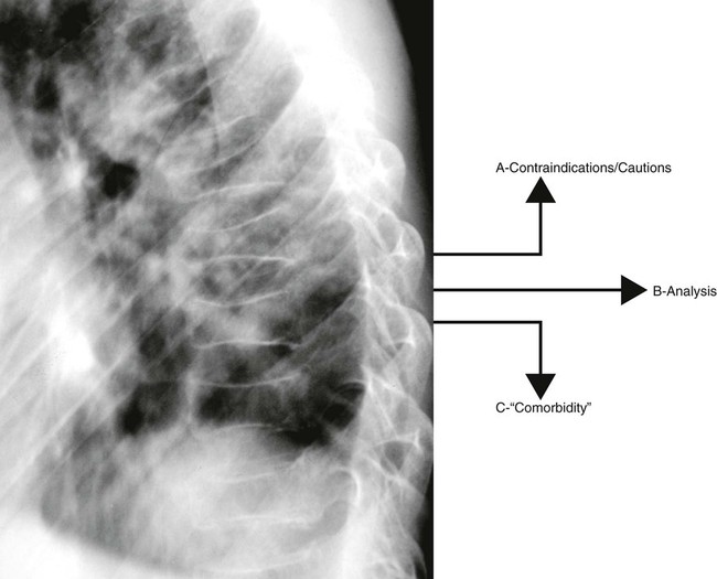
In chiropractic practice, radiographs are generally considered a standard first-step imaging protocol when evaluating degenerative and inflammatory joint disease, fractures, infections, and neoplasms.26 The hands-on management approach of chiropractors demands attention to biomechanical influences and potential structural contraindications for intended interventions (Fig. 5-1). Nonchiropractic clinicians, whose management of low back pain centers on exercise, patient education, pharmaceuticals, and other clinician-passive therapies, have less use for the biomechanical or structural information obtained from radiographs. Therefore, these practitioners can easily adopt a more conservative approach to taking radiographs for musculoskeletal spine complaints than can chiropractors or other practitioners, who apply manual intervention.

image
FIG 5-1 Three streams of information from the radiograph. In the context of delivering chiropractic care (or other manual approaches to patient care), radiographs provide three types of information: (a) information related to possible structural contraindications or cautions to delivering a corrective force into the demonstrated anatomy; (b) information that may influence the direction or technical approach to delivering a corrective force; and (c) information related to diseases, conditions, or findings that are seen in addition to the chiropractic subluxation, osteopathic lesion, or primary reasons the radiographs were taken. For instance, this radiograph demonstrates osteopenia that will influence the delivery of the chiropractic adjustment (stream A). There may be biomechanical or degenerative features that will cause the chiropractor to adjust one segment over another, thereby influencing the technical approach to the patient (stream B). Last, the compression fractures may result from an underlying aggressive pathology, such as metastasis, which would be of great concern to the clinician, beyond the issue related to the chiropractic subluxation or the initial concerns of the clinician (stream C).

Many radiographs taken in a chiropractic setting are interpreted as normal for serious bone pathology,27 but may contain biomechanical or structural information allowing the chiropractor to be more successful with technical aspects of formulating and applying the patient’s management plan. However, more research into the reliability, validity, and clinical usefulness of biomechanical and structural information gleaned from radiographs is necessary. Also, evidence is needed to clearly justify the added costs, define criteria of patient selection, and facilitate advancements in care delivery.

All clinicians, regardless of therapeutic approach, are concerned with serious pathology appearing as routine low back pain. Clinical red flags suggesting the presence of serious pathology have been developed and are helpful for directing patient selection (Box 5-1). In the absence of these red flags, significant spinal pathology is estimated in only 1 of 2500 patients.28 Deyo and Diehl29 evaluated 1975 walk-in patients at a public hospital to estimate the prevalence of cancer as an underlying cause of patient back pain. Using an algorithm that generally reflects the questions listed in Box 5-1, only 22% of these patients would have received x-rays; this proportion includes all those who were later found to have cancer.

Box 5-1

Clinical Red Flags of Serious Spinal Pathology (e.g., Infection, Tumor, or Fracture)

Unexplained weight loss

Personal history of cancer

Unexplained fever

Age greater than 50 years

Intravenous drug use

Prolonged corticosteroid use

Severe, unremitting pain at night

Trauma sufficient to cause fracture or injury

Pain that worsens when the patient is lying down

Features of cauda equina syndrome

Urinary retention

Bilateral neurologic signs or symptoms

Saddle anesthesia

Data from Deyo RA, Diehl AK: Lumbar spine films in primary care: current use and effects of selective ordering criteria, J Gen Intern Med 1:20, 1986.

It should not be assumed that instituting guidelines, such as those listed in Box 5-1, will lead to less use. For example, Canadian researchers found that if the guidelines listed in Box 5-1 had been applied to their study population of 963 patients in a private medical family practice setting, 44% would have undergone radiography, increasing actual use by 238%. Considering patient follow-up, these researchers concluded that the sensitivity of the guidelines to detect fractures and tumors was higher than the physicians’ use patterns, but their specificity and positive predictive values were low.30

Parallel and similarly controversial issues surround the application of specialized imaging (e.g., whether MRI should be ordered for a patient in whom a disc herniation is clinically suspected yet neurologic findings are limited). This is especially true when approximately 25% of normal adults demonstrate acquired spinal stenosis and 33% have a disc herniation,31 two key MRI findings.

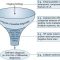
In the absence of clear guidelines, clinicians must adhere to a logical rule for the use of all diagnostic procedures: If the patient’s diagnosis or management is likely to significantly change from information routinely provided by the diagnostic procedure in question, then the study should be performed; if the necessary information is not routinely provided by the procedure or knowing the information obtained will not change the patient’s management, then the procedure should not be performed. This basic rule can be elaborated upon by an evidence-based approach to the decision. Similar to evidenced-based clinical practice generally, evidence-based radiology is an important approach to guide the practice of radiology. The clinician is encouraged to identify existing evidence in a systematic fashion, assimilate the information found in the context of the clinical question, and make a critical review of the strength of the evidence, and to recognize that the lack of evidence is not equivalent to negative evidence. Therefore, considerations of standards of practice and practitioner experience are important also.

Clinical decisions on radiology are then based on the best current evidence, clinical experience, and patient values.32 The information in Box 5-2 patterns the thought process behind an evidence-based approach through the presentation of a clinical decision rule.

Box 5-2   Anatomy of a Clinical Decision Rule

Clinical Scenario

A 35-year-old patient comes into the office explaining a recent history twisting his ankle. There is swelling and ecchymosis visible during physical examination. The patient is able to bear weight on his ankle by standing and there is no particular tenderness on the medial or lateral malleoli following mild clinician palpation. The navicular and the base of the fifth metatarsal are not tender to touch. Given the patient’s presentation, the clinician is reasonably confident that the injury is a sprain and not a fracture.

Clinical Decision Rule

The clinician’s specific conclusion in the scenario above is based on the application of the Ottawa Ankle Rules (OAR). The OAR are clinical decision or clinical prediction rules based on evidence and clinical experience, incorporating patient values to receive the best care possible. These are different from guidelines in that clinical decision rules provide estimates of probability of a disorder and are created through a process of derivation, validation, and impact analysis. Guidelines are created through a best evidence synthesis with a consensus of experts, and are not necessarily directly focused on clinical outcome prediction. Specifically, the OAR holds that radiographic examination of an injured ankle is indicated to evaluate for suspected fracture if there is malleolar region pain and any of the following findings: bone tenderness at the posterior margin of the medial or lateral malleolus or the inability to bear weight for four walking steps. Also, radiology of the foot is indicated to assess for fracture if a patient has pain in the midfoot zone and any of the following findings: bone tenderness at base of the fifth metatarsal or the navicular or the inability to bear weight for four walking steps.

The OAR were formed from a multistep process that can be applied to create clinical decision rules for a variety of applications. The steps are summarized as the following:

Step 1—Derivation. Potential predictors of a condition or disorder are identified and assessed in light of a gold standard diagnostic procedure. For example, related to the OAR, Stiell et al. published a study in 1992 that identified 32 variables that were assessed for association with radiographically diagnosed ankle fracture.33 Using statistical measures of association these variables were refined to create clinical decision rules.

Step 2—Validation. Validation occurs is multiple stages. Narrow validation occurs when the rule is tested in a population similar to that used for the derivation. Broad validation occurs when the rule is tested in a variety of settings with different patient populations. For example, related to the OAR, a 1993 study prospectively evaluated clinical decision rules in two stages. In the first stage more than 1000 adults with acute ankle injuries in two university emergency departments were assessed using the derived rules. Using the data from the first stage the rules were refined and applied to 453 patients in the second stage. This study demonstrated 100% sensitivity of the rules for identifying ankle and foot fractures and provided a narrow validation.33 Further, a 2003 systematic review published in the British Medical Journal pooled the results of 27 studies involving different settings and different patient populations. More than 15,000 patients were used in the meta-analysis supporting the accuracy of the clinical decision rule. This provided broad validation.34

Step 3—Impact Analysis. Impact analysis is necessary to determine if the rule will actually change clinician behavior and provide definite benefit, either financial or in patient outcomes. A clinical decision rule that is wieldy or difficult to apply will not be adopted unless there is a highly significant benefit. For example, related to the OAR, a large multicenter trial involving community and teaching hospitals in Canada compared outcomes from 1 year prior to implementing the ankle clinical decision rules to the following year. More than 12,000 patients and 200 clinicians were involved. There was a significant reduction in x-rays with a corresponding reduction in patient cost. This study had high compliance by physicians who accurately applied the rules.33

In summary, the degree to which a clinician can rely on a clinical decision rule is dependent on the level of evidence supporting its use. To provide some guidance, rules may be placed into four categories to aid the clinician’s selection.35

Level I. At least one prospective validation study has been done in a population different from the derivation group. At least one impact study demonstrating usability and benefit was done. As always, the clinician should evaluate the strength of these studies. These clinical decision rules can be utilized with a high degree of confidence by the clinician. The Ottawa ankle rules fall into this category.

Level II. A least one well-designed validation study has been performed with a spectrum of patients and a number of clinicians providing broad validation. In this circumstance, no impact analysis has been performed.

Level III. Narrow validation has been accomplished in a patient population not significantly different from the derivation population.

Level IV. The rule has been derived but no prospective validations studies have been performed.

Image Interpretation

Equipment and Resources

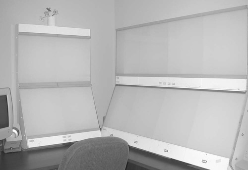
Before the skills and knowledge of the interpreter can be brought to task, the images should be clearly displayed and reference material should be close at hand. This is a digital age, but clearly the revolution has not permeated all segments of the population evenly. Although many clinics have moved to filmless methods for acquiring and displaying images, not all are as advanced. Much radiology, particularly plain film, is still accomplished traditionally; this produces radiographs that should be viewed on illuminated light boxes. These view boxes generally are available in two sizes, the standard 14 × 17-inch view box, and a larger 14 × 36-inch view box that accommodates a full spine radiograph, the type often used to assess scoliosis. Standard 14 ×17-inch view boxes are combined in various configurations to create a viewing station (Fig. 5-2).

image
FIG 5-2 View box station. View boxes are arranged in various formats to construct a viewing station. This picture exhibits a simple four-over-four bank on the right and a two-over-two bank to the left of it.

High-volume centers may invest in a viewing system with rotating panels or belts that pass the films in front of a stationary bank of lights, because placing the films on and off the view box can consume a considerable amount of time. This system allows many cases to be stored and viewed quickly, without the need to shuffle through the films of each case as they are put on and off the view box.

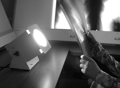
A “hot” or “bright” light is another important tool necessary for film interpretation (Fig. 5-3). The hot light produces a controllable high-intensity beam of light that helps the interpreter view the overexposed (dark or radiolucent) areas of the film. The intensity of the light can be controlled with a pedal that allows the interpreter to match the brightness of the light to the darkness of the radiograph. Even radiographs that are executed under the highest technical standard have regions of overexposed anatomy. Some of the more common and significant pathologies often hide in the overexposed areas of the film, making it difficult to recognize them when viewing the films only on a view box. Therefore, a hot light is an essential tool to a thorough film interpretation.

image
FIG 5-3 Hot light. The hot light (also known as a bright light) is an essential tool to film interpretation. By using a hot light, the interpreter is able to view the overexposed regions of the film. Some of the most serious pathologies (e.g., lung nodules, aneurysms) are common to the overexposed regions of a radiograph and are more easily seen with the aid of a hot light.

It has been said that “a radiologist without a ruler is a radiologist in trouble.”36 Although the sentiment underscores the importance of clinical intuition, observation, and training, the reality is that handy access to rulers, protractors, or other measuring devices allows more accurate quantification of structural abnormalities. For instance, the degree of spondylolisthesis is related to the likelihood of its further progression, the rate of growth of a pulmonary nodule predicts its malignant potential, the degree of scoliosis is central to the management of the case, and so on.

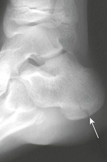
Reference texts should be close at hand. The usefulness of some radiology books transcend the typical, such as Keats’ Atlas of Normal Roentgen Variants That May Simulate Disease.37 A recent edition of Keats’ atlas should be close to the reading area. This book is a regional atlas of abnormal film findings that are normal variants of anatomy. It is comprehensive and includes both subtle and grossly abnormal cases. Recognizing that an abnormal finding is a normal variant saves time and examination costs related to erroneous additional evaluation. For example, view the case exhibited in Figure 5-4 of a 12-year-old with a history of trauma. The calcaneus clearly looks fractured, but the radiolucent defect actually represents an unfused secondary growth at the center of the calcaneal tuberosity. A similar case is noted in the third edition of Keats’ book.37 Recognizing that this is a normal variant and not a fracture ensures that time and expense are not wasted, nor that treatment is inappropriately provided.

image
FIG 5-4 Lateral ankle of a 12-year-old who recently suffered a trauma to the calcaneus. The radiolucent line appearing as a fracture (arrow) actually represents a normal appearance of the secondary growth centers. This case demonstrates how closely some normal findings and variants of normal may simulate a disease state. Courtesy C. Robert Tatum, Davenport, IA.

Errors in Film Interpretation

Interpreter error may arise from a failure to see, recognize, or understand the significance of a lesion. Although error rates of 20% to 30% have been reported,38 the more contemporary literature indicates that approximately 1% to 3% of plain film interpretations done by nonradiologists contain important errors. Most of these studies compare the film interpretations of attending emergency department physicians with planned overread interpretations of radiologists.39Seltzer40 estimates that 8% of interpretations by medical radiology residents contain potentially clinically important errors, which suggests that misinterpretation is increased in interpreters with fewer qualifications. Complex studies (e.g., CT)41 and studies on pediatric patients42 also increase misinterpretation. Fractures are the most often-missed lesions.4345 Training is associated with more accurate interpretation,46,47 but radiologists are not immune to misinterpretation, as studied with various methods and imaging modalities.4853 Most studies focus on false-negative readings; however, false-positive readings occur among radiologists43 and nonradiologists54 alike. False-positives promote continued patient evaluation, adding to cost, which is estimated to average $85 per false-positive reading.54 Unnecessary examinations also result in increased risk of complications associated with imaging.

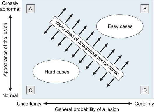
Alleged diagnostic errors account for the majority of legal cases related to radiology departments.55 However, not every missed lesion constitutes evidence of negligence. Statistics pointing to related rates of missed lesions, limitations of normal human visual perception, image quality, and many other factors influence image interpretation, and may be mitigating factors for image misinterpretation.5562 The conceptual difference between errors in interpretation and those arising from perceptual variations is well described by Robinson.44 The former assumes the diagnosis is known and generally agreed upon as a lesion; the latter does not (Fig. 5-5).

image
FIG 5-5 Errors in image interpretation. This figure depicts a relationship between the imaging appearance of a lesion (size, shape, etc.) and the probability that the apparent lesion truly is one. A, At times, there may be an uncertain or mixed interpretation of a grossly abnormal feature (e.g., importance of a lumbosacral transitional segmentation or spondylolisthesis). B, Easy cases to interpret are defined as those that appear clearly abnormal on imaging and have a high degree of certainty that they are real lesions. Misinterpreting such a case is a clear error. C, In contrast, difficult cases are those that present with only mild departures from the appearance of normal anatomy and are associated with doubtful conclusions on whether a true lesion is present. Misinterpreting this case may reflect a genuine difference of opinion among experts, representing variation in opinion more than direct error. D, Last, only a small deviation from normal anatomy may correlate to a certain lesion. For example, a small corner fracture of the phalanx appears subtle, but has a certain interpretation as a lesion. The watershed of acceptable performance represents the line between clear-cut error and the inevitable difference in opinion existing between and among professionals. From Robinson PJ: Radiology’s Achilles’ heel: error and variation in the interpretation of the Röntgen image, Br J Radiol 70:1085, 1997.

The goal of film interpretation is to eliminate as many misinterpretations as possible. There is a substantial literature addressing the topic of radiologic interpretation (Table 5-1). The literature and conventional wisdom indicate that although it is impossible to eliminate human error, and therefore mistakes of radiologic interpretation, attention to common principles should prove beneficial (Box 5-3).

Box 5-3   Suggestions to Reduce Radiologic Interpretation Errors

1. Become familiar with the patient data. The age, gender, and ethnicity of the patient may offer potent predictors of what disease process is represented on the radiographs. For instance, a 2-cm solitary radiodense defect of the L4 vertebral body is likely a bone island in a patient younger than 30 years of age. By contrast, a metastatic deposit needs to be excluded in a patient older than 40 years of age. Patient demographics directly influence clinical decision making.

2. Become familiar with the clinical context of the study. The clinical rationale for the study should be known before a patient’s images are viewed. The interpreter should have a good idea of what he or she will encounter before the films come out of the processor. For example, are the films done to evaluate the patient for a clinically suspected rib fracture? Knowing the clinical context is essential to a thorough interpretation.

3. Assess technical factors, image quality, and artifacts. The images should be of sufficient quality and the correct area of clinical interest. Film interpretation is directly and negatively influenced by poor technical factors, such as improper patient positioning, patient motion, and many others problems that are more fully discussed in Chapter 1. Faint shadows of pathology may not be visible if the images are underexposed or overexposed. Patient motion is probably the most common technical defect that degrades image interpretation. Also, all of the clinically relevant anatomy must be visible to the degree expected. Images taken when the patient is recumbent may cause some of the anatomy to appear quite different. A case in point is the difference in size of the heart shadow on recumbent and upright films, or inspiration versus expiration patient instructions on upright films. Clothing and other artifacts can form ambiguous presentations on radiographs.

4. Search the images using an intentional, thorough visual path. Image interpretation has a greater chance of being successful if steps are taken to view the entire image in a complete manner. One popular approach to film interpretation involves a review of the alignments (A) of structures, bone (B) elements, cartilage (C) or joint spaces, and soft tissues (S). This is known as the “ABCS” approach to film interpretation and the method ensures that all of the anatomy is viewed completely. However, the interpreter should not be a slave to the ABCS sequence of film interpretation. For some, the ABCS approach may be more naturally applied as a BCAS or CABS sequence. The important point is that all of the anatomy is viewed; the order is less critical.

    When an abnormal finding is discovered, a free search path is invoked to follow the features of the disease. Once all of the related findings have been observed, the interpreter returns to a fixed sequence to ensure that all of the anatomy has been viewed. The search path never should be haphazard. It needs to intentionally follow the predetermined path of the anatomy (e.g., ABCS) or trail of pathologic findings (e.g., observed fracture line, associated angulation, soft-tissue distension).

5. Patients are entitled to more than one problem. As mentioned, the radiographs should be searched completely for defects in the ABCS. It is especially important to be vigilant to a complete search pattern after a lesion is uncovered. For instance, at times the interpreter may become so preoccupied with the first abnormality detected that a second or third lesion may be overlooked. This is a well-established phenomenon known as a “satisfaction of search error.”82,91 By definition, satisfaction of search errors represent omissions of underreading images. The satisfaction of search phenomenon is more likely to manifest when the first lesion is generally more attention-grabbing than the subsequent, more subtle lesion.92

    Although the causes of satisfaction of search errors are not explicitly defined, these errors may be reduced if interpreters are aware of the tendency to miss subsequent lesions, and hopefully develop a tendency to closely look for a second lesion whenever a first lesion is found, a third when a second is found, and so on until the anatomy is viewed completely. A complete search pattern is the only defense to avoid this well-known pitfall of image interpretation.

6. Compare what is seen with the “mind’s eye of normal.” After a review of the patient’s demographics, the rationale for examination, and technical issues related to the image, image interpretation next begins with a thorough search of the displayed anatomy for any deviations from normal. This step is the most crucial of the film interpretation process. Image interpretation requires an excellent knowledge of normal anatomy. Abnormality quickly catches the eye when normal is understood.93

7. Common things are commonly seen (or, rare things are rarely seen). This concept underscores the importance of trying to explain the cause of abnormal findings by starting with the common pathologies and working to the less common differentials. For instance, a fragmented, radiodense, small proximal epiphysis appearing on hip radiographs of a 6-year-old boy could signify hypothyroidism, but there is a better chance that it represents traumatically induced avascular necrosis.

8. Be proactive, not reactive. Excluding common pathologic presentations should be proactively attempted; that is, pathologies that are common to some radiographs should be routinely investigated. For example, when an anteroposterior (AP) open-mouth projection is viewed, an odontoid fracture should specifically be looked for. Signs of an aneurysm of the abdominal aorta on a lateral lumbar projection, a femoral neck fracture on an AP view of the pelvis, and so on should be checked. Features of the pathology should not simply be reacted to; common pathologic presentations should be proactively eliminated, especially those suggested clinically.

9. There is no substitute for experience. Critically interpreting large numbers of radiographs will help interpreters develop a strong sense of normal anatomy, and allow subtle abnormal shadows to be more apparent.

10. Consult with someone on difficult or ambiguous cases. The social literature says that two minds are better than one. A group decision generally is more accurate than an individual conclusion. Keeping with this theme, when ambiguous findings are recognized or intuitively suspected, it may be helpful to obtain a second opinion to resolve any controversy and arrive at a valid film interpretation. Interestingly, research suggests that when asking another interpreter to view the film, it is better to blind the second interpreter to the suspicious areas of the film found by the first examiner. Swenson and Theodore94 found that second interpreters of chest films were more accurate if they read the films using a “free search pattern,” unencumbered and independent of the exact prior concerns of the first interpreter. The theory that supports this assertion is described as “superiority of search.” The theory holds that when interpreters review standard radiographic views (e.g., posteroanterior [PA] chest film), they go through a process of skilled perceptual filtering that allows them to recognize abnormal findings. Diagnostic radiology is a visual interpretation reliant on knowledge and visual acuity. It is dependent on the ability to sort information to arrive at clinically meaningful conclusions. Moreover, if the search pattern used by the radiologist is interrupted (in this case, by being tipped off to what to look at), the perceptual mechanisms are bypassed, resulting in a less accurate interpretation.94 There is empiric evidence that second opinions are helpful to the interpretation of chest radiographs,47,95 barium enemas,84 and mammography.10,57,61

11. Search for links between findings and various views. Cognitively linking related findings together should be attempted in hopes of developing a perceptional flow to the image interpretation. Triangulation between the available views of the region (e.g., PA and lateral chest or AP, AP open-mouth, lateral cervical spine) should be used. Some interpreters embrace the concept that related anatomy should be reviewed together. For instance, when viewing a PA chest film it makes more sense to view all of the ribs separate from viewing the pulmonary tissues, as opposed to viewing the first rib and estimated pulmonary tissues concurrently.

12. Eliminate extraneous light. The ambient room light should be low, and view boxes that are not displaying images should be turned off.

13. Compare current findings with those on past radiographs. Past imaging studies are very useful to aid in the interpretation of current radiographs.16,34 Berbaum found that normal comparison images were especially helpful for those interpreters who were in the earlier stage of training.34 This is believed to be related to the perceptual operation in which single perceptions from the old and new film combine to form a common, third, unique perception. Comparison with past radiographs also helps to document the progression of a lesion. The stability of a lesion over time is a key predictor of its aggressiveness. For example, a 1-cm lung nodule not seen on radiographs 6 months previously likely indicates a malignant etiology of the nodule. If the nodule was on an early film and of consistent size on the past images, an etiology of granulomatous infection is likely to explain the nodule. However, caution is necessary. At times the progression of a pathology may be so subtle as to be missed when very recent past radiographs are viewed. Use of multiple comparisons, including old past images, is best to avoid this pitfall.

14. The problem is perception. Image interpretation is a function of perception more than visual acuity. The question is not what can be seen, but identifying what is seen. Interpreters tend to overlay personal bias onto the process of image interpretation. It takes discipline to focus on the objective interpretation of the demonstrated anatomy. Perception is made better by experience and continually correlating what is seen to what is actually present. This is learned best by iterations of comparing one’s interpretation with that of someone more proficient.

15. Is the abnormal finding real? When abnormal shadows are seen on the image, it should first be considered that they may represent nothing more than a presentation of normal anatomy, artifact, or confluence of overlying shadows. It is common for superimposed structures to form a resultant shape. For instance, the pulmonary arteries coursing in multiple directions across the lung may combine to give the appearance of a circular, cystic pulmonary defect. The resulting “virtual image” is termed a subjective image. It is not real; rather, it is an illusion conjured in the mind of the interpreter.

16. Give special attention to the problem areas of the radiograph. Some areas on the film are more likely to contain pathology than others. For instance, attention must be paid to interpret the lung apices on the AP lower cervical radiograph; the atlantodental interval and sella turcica are important areas that are often neglected on the lateral radiograph. Problem areas for various projections are listed in Figures 5-12 to 5-20. Generally, special attention should be given to the overexposed, radiolucent areas of all projections as common sites for pathology.

17. Be organized. Successful film interpretation requires some attention to detail. The patient’s images should be accompanied by a correlating history and pertinent clinical data, inclusive of any past imaging studies that may assist the interpretation. Resources (e.g., books, rulers, protractors, voice recorders, alcohol to erase pencil lines, dry erase markers for annotating abnormal findings) should be close at hand. Part of being organized involves consistency in how the images are assembled on the view boxes. For instance, many interpreters feel compelled to place the lateral radiographs on the view box so the patients are facing to the interpreter’s reading left, or view the films in a consistent sequence: lateral…AP…oblique, etc.

18. Attention to environment. The film reading environment should be quiet and free of distractions. Maintaining low ambient room light and turning off view boxes without films are both empirically associated with successful image interpretation.

19. What is the clinical impact? The purpose of diagnostic imaging is to gather data to assist clinical decision making. Therefore all abnormal findings should be interpreted in light of their clinical significance with appropriate follow-up imaging or procedures formulated into the report generated from the images.

20. Do not ignore intuition. The largest portion of the variation in film interpretation is unexplained. Sometimes the only predictor of an abnormality is that lingering sense that something is being missed. This feeling often drives extra attention that may eventually reveal a subtle defect. Perhaps the eyes are seeing something that the brain cannot immediately comprehend.

21. Resist overinterpreting the study. No matter how closely they are scrutinized, plain film radiographs do not reliably detail a disc lesion, ependymoma, hydrosyringomyelia, or a host of other defects. The clinical utility of the imaging modality must always be matched to the clinical question, and the modality’s limitations must be considered. The temptation must be resisted to assume that the patient’s problem is illustrated on the imaging study being reviewed. Also, a link must not be assumed between the patient’s clinical problem and radiographic abnormalities that happen to be present. The early literature is replete with assumptive correlations that could not be supported when investigated in greater depth.

22. Confirm findings with other views or studies. If findings are equivocal, further views, contrast, specialized imaging, and so on should be used to increase the certainty about something found.

TABLE 5-1

ANNOTATED BIBLIOGRAPHY OF SELECTED ARTICLES ADDRESSING RADIOLOGIC IMAGE INTERPRETATION

Only gold members can continue reading. Log In or Register to continue

Topic Article Summary
Ambient light Alter AJ et al.65 Employing low ambient room light, illuminating only films being viewed, and masking the radiograph around areas of interest improves visual performance; however, is cumbersome to implement completely in a clinical setting.
Appearance of the lesion Krupinski EA et al.66 This study found that physical features of pulmonary nodules do not attract attention as measured by “first-hit” fixation of the interpreter’s gaze; however, certain features do tend to hold the attention once the nodule has been fixated. The combination of all features influences whether or not it is detected.
Experience Herman PG et al.48 After having several interpreters view a series of chest radiographs, the authors found that, “Once an individual’s radiology education has progressed beyond a fundamental level, individual reader characteristics overshadow experience (and training) in the accuracy of chest film interpretation.”
Experience

Stay updated, free articles. Join our Telegram channel

Feb 2, 2016 | Posted by in RESPIRATORY IMAGING | Comments Off on Film Interpretation and Report Writing

Full access? Get Clinical Tree

Get Clinical Tree app for offline access