Gastric Volvulus
Ho Chia Ming
Ellie R. Lee
CLINICAL HISTORY
50-year-old patient with 48 hours of worsening left-sided chest pain, persistent nausea, and vomiting.
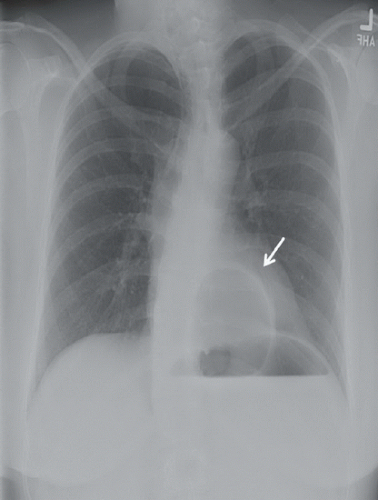 FIGURE 95A |
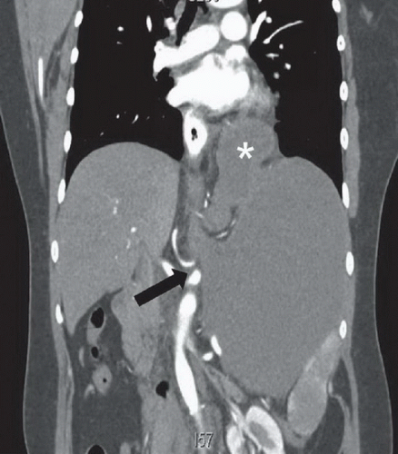 FIGURE 95B |
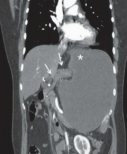 FIGURE 95C |
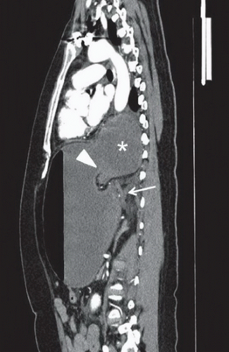 FIGURE 95D |
FINDINGS
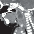
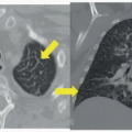
Figure 95A: Plain radiograph of the chest shows a retrocardiac air-filled mass, corresponding to the gastric antrum above the diaphragm (arrow). Figure 95B: Coronal contrast-enhanced CT image of the upper abdomen demonstrates a distended stomach with abnormal orientation. The gastroesophageal (GE) junction (black arrow) lies inferior to the left diaphragm and below the gastric antrum (asterisk), representing a mesenteroaxial volvulus. The greater curvature remains to the left of the lesser curvature. Figure 95C: Coronal CT image of the upper abdomen anterior to Figure 95B demonstrates the gastric antrum (asterisk) in the left upper abdomen and nondilated proximal duodenum (arrows) superior to the gastric fundus. Figure 95D: Sagittal CT image of the abdomen shows herniation of the dilated gastric antrum (asterisk) into the thorax. The gastric antrum and pylorus (arrowhead) is located superior to the GE junction (arrow), compatible with a mesenteroaxial volvulus. Gastric fluid-filled distention was related to gastric outlet obstruction.
Related posts:


Stay updated, free articles. Join our Telegram channel

Full access? Get Clinical Tree


