Types of bowel surgeries, their indications, and postprocedural anatomy
Esophagectomy
All esophagectomy techniques involve partial or complete resection of the esophagus with a new anastomosis to esophagus, stomach, or bowel. Benign indications include esophageal perforation (iatrogenic from endoscopy/biopsy/balloon dilation, traumatic or self-induced from Boerhaave syndrome), refractory strictures from peptic ulcers/radiation/caustic ingestion, and esophageal fistulas (congenital or iatrogenic from trachea or bronchi). The most common malignant causes include adenocarcinoma and squamous cell carcinoma of the esophagus.
- ■
Right-sided transthoracic esophagectomy is preferred when the upper two-thirds of the esophagus is involved given the aorta would otherwise limit access, whereas a left-sided approach can be used when the distal esophagus is involved.
- ■
The Ivor-Lewis technique is best for pathology of the mid-esophagus and combines a laparotomy with right thoracotomy and intrathoracic anastomosis ( Fig. 7.1 ).
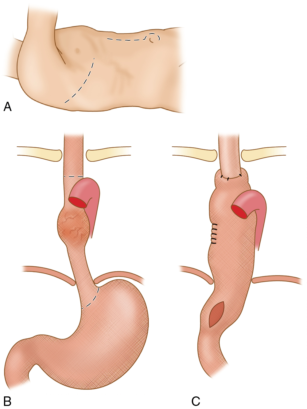
Fig. 7.1
Overview of right thoracotomy (A) with esophageal resection, (B) gastric mobilization, and (C) intrathoracic anastomosis.
(From Sahani DV, Samir AE. Abdominal Imaging , ed 2. Philadelphia: Elsevier; 2017.)
- ■
Transhiatal esophagectomy was developed because of complications from the transthoracic approach in treating long segment pathology. It involves mobilizing the esophagus through the esophageal hiatus, after which the entire esophagus is transected and the stomach is pulled up and anastomosed to the cervical esophageal remnant ( Fig. 7.2 ).
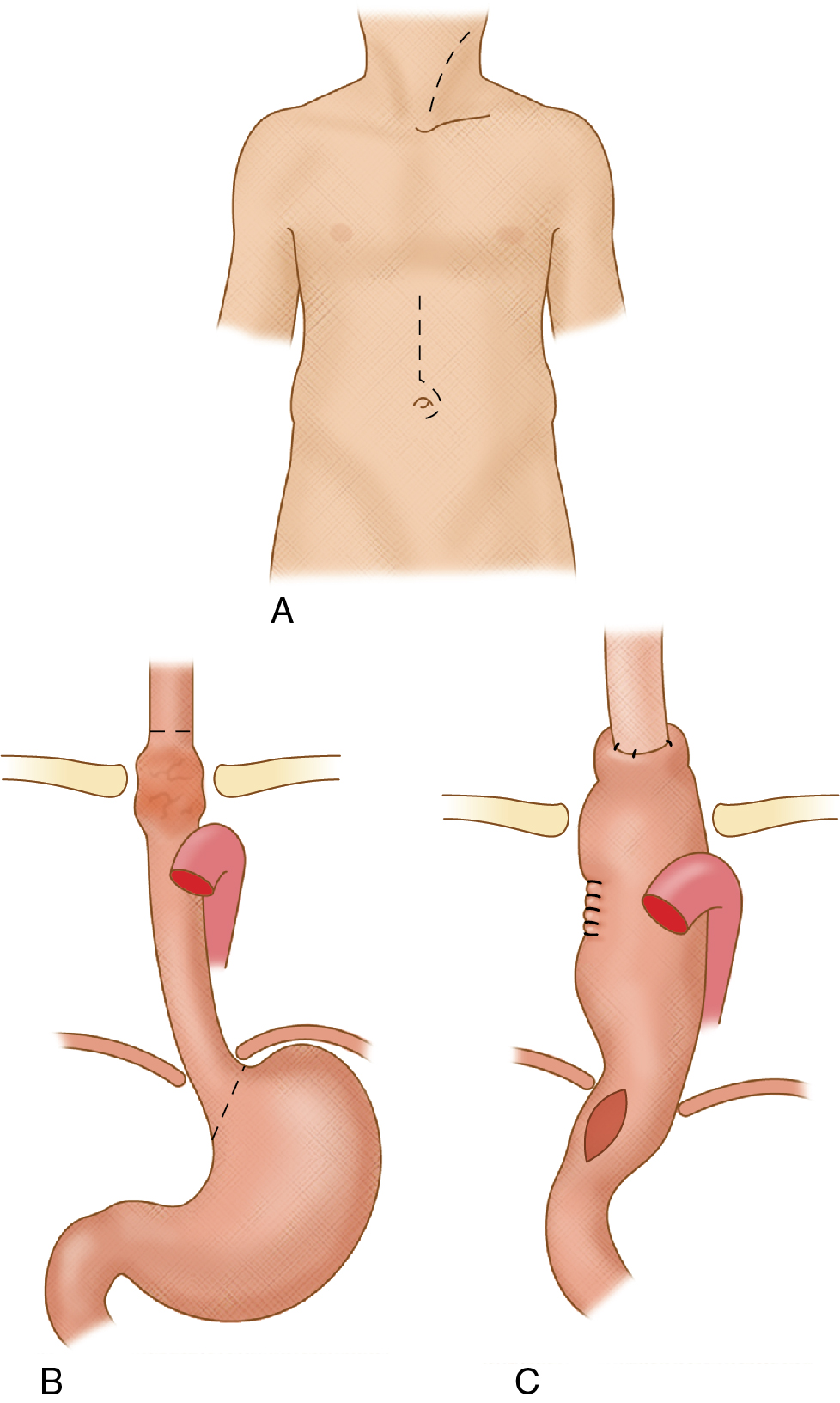
Fig. 7.2
Overview of transhiatal esophagectomy (A) with gastric mobilization and gastric pull-up (B) for cervicoesophagogastric anastomosis (C).
(From Sahani DV, Samir AE. Abdominal Imaging , ed 2. Philadelphia: Elsevier; 2017.)
Antireflux surgery
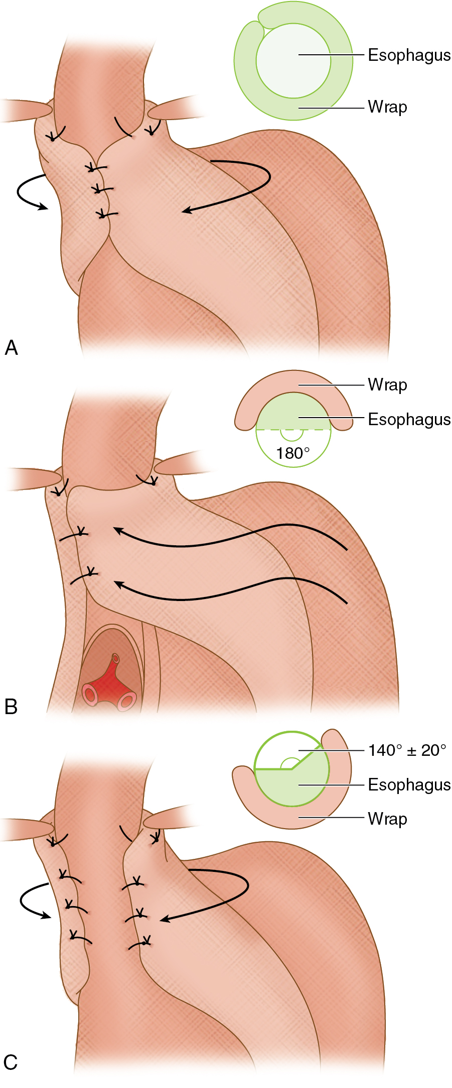
The idea behind antireflux surgery is to create a valve-mechanism to reestablish gastroesophageal junction competence ( Fig. 7.3 ). When associated with a sliding or paraesophageal hernia greater than 5 cm, this too will need to be corrected by approximating the pillars of the diaphragm crura posterior and inferior to the esophagus. In more severe cases, an anterior abdominal wall gastropexy can be performed to reduce the risk of hernia recurrence. Indications for antireflux surgery include gastroesophageal reflux disease refractory to medical treatment, severe esophagitis by endoscopy, benign stricture, or early Barrett esophagus without concerning features. Relative contraindications include severe esophageal dysmotility and achalasia. The most common techniques require a normal length esophagus, and the technique of choice then depends on the patient’s esophageal motility and gastric emptying (the fundus and upper body of the stomach are responsible for gastric emptying, and underlying gastroparesis may worsen with fundoplication, which may be prevented with concurrent pyloroplasty at the time of surgery).
- ■
The Nissen fundoplication is preferred when the esophagus and stomach are normal in motility, and can be performed via laparotomy or laparoscopically. It uses the stomach fundus and wraps it around the distal esophagus 360 degrees before being sutured in place.
- ■
Partial (i.e., Toupet) fundoplication techniques are best when there are underlying issues with esophageal and/or gastric motility. These procedures incompletely wrap the fundus around the distal esophagus to avoid worsening the underlying esophageal/gastric motility but at the same time provide some degree of reflux prevention. As such, these do not perform as well long term and have higher rates of reflux recurrence.
Bariatric surgery
- ■
Bariatric surgeries are typically reserved for the morbidly obese (body mass index >40 kg/m 2 , or 35 kg/m 2 with comorbidities) to improve quality of life and comorbidities (such as heart disease, sleep apnea, and diabetes). Restrictive procedures, such as vertical banded gastropexy, gastric sleeve, and gastric banding serve to reduce caloric intake by limiting gastric capacity. Malabsorptive procedures, such as jejunoileal bypass and biliopancreatic diversion with duodenal switch reduce the absorption of calories by decreasing the length of the small intestine. Roux-en-Y gastric bypass combines both principles by limiting caloric intake with a gastric pouch and reducing absorption through a gastrojejunal component.
- ■
Roux-en-Y gastric bypass is typically the preferred method owing to decreased hospital stays and faster recovery. Preoperative planning for this procedure must include exclusion of underlying malignancy or inflammatory bowel disease (IBD), as well as confirming normal anatomy of the proximal gastrointestinal (GI) tract via fluoroscopy (i.e., an upper GI study). For postoperative imaging purposes, the four suture lines to consider are that of the gastric pouch (when the fundus is transected), the gastrojejunal anastomosis to the gastric pouch, the transected fundus of the remaining stomach, and the jejunojejunostomy. The Y-limb (i.e., afferent or pancreaticobiliary limb) consists of the excluded stomach, duodenum, and proximal jejunum ending at the jejunojejunostomy, and the Roux-limb (i.e., efferent or antegrade limb) includes the gastric pouch and anastomosed jejunum to the level of the jejunojejunostomy ( Fig. 7.4 ).
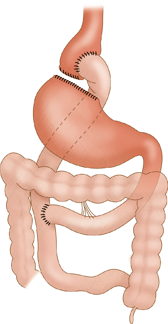
Fig. 7.4
Roux-en-Y gastric bypass.
(Modified from Cameron JL. Current Surgical Therapy , ed 7. St Louis: Mosby; 2005: p. 99.)
- ■
Gastric surgery
Gastric surgeries are performed for both benign and malignant conditions. The location of the pathology will usually dictate which type of surgery is best.
- ■
For small tumors or severe ulcerative disease in the distal two-thirds of the stomach, a partial gastrectomy (i.e., antrectomy) may be a viable option; either a Billroth I or II reanastomosis is commonly used to restore the enteric anatomy. The Billroth I procedure involves an antrectomy and an end-to-end anastomosis between the remnant stomach and the duodenum. In the Billroth II operation, after the antrectomy is performed, the duodenal stump is closed and a gastrojejunal anastomosis or a side-to-side duodenojejunal anastomosis is created.
- ■
Total gastrectomy is typically reserved for severe and diffuse ulcerative disease with complications (perforation or strictures), for tumors in the proximal third of the stomach or which are infiltrative, or for large mid-gastric tumors. For these cases, a Roux-en-Y esophagojejunostomy with end-to-side anastomosis is created, with the Y-limb including the duodenum and proximal jejunum and the Roux-limb beginning at the esophagojejunal anastomosis and continuing distally ( Fig. 7.5 ).
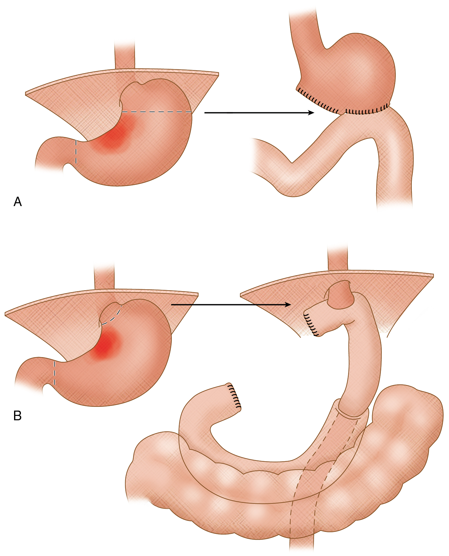
Fig. 7.5
A, Total gastrectomy with a Roux-en-Y anastomosis. B, Subtotal gastrectomy with a Billroth II anastomosis.
(Modified from Townsend CM. Sabiston Textbook of Surgery , ed 17. Philadelphia: Saunders; 2004: p. 1310.)
Surgery for pancreatic pathology
For patients who present with a malignancy of the pancreatic head (classically adenocarcinoma, less commonly metastases, or cholangiocarcinoma), patients who require management of pancreatic or duodenal trauma, or for refractory cases of chronic pancreatitis, a Whipple procedure (also known as a pancreaticoduodenectomy) can be performed to either extend the patient’s life expectancy or improve quality of life. If performed because of a malignancy, potential contraindications to surgery include whether or not there is more than 180-degree tumor encasement of important adjacent structures (such as the celiac trunk, superior mesenteric artery, superior mesenteric vein, or inferior mesenteric vein), or the presence of distant metastatic disease.
- ■
The procedure itself (most commonly) involves resection of the gastric antrum, entire duodenum, the pancreatic head, common bile duct, gallbladder, and first 15 cm of jejunum. From there, the new anastomoses that are reconstructed include a gastrojejunostomy (involving mid-jejunum), a pancreaticojejunostomy (end-to-side anastomosis with the proximal jejunal stump), and a choledochojejunostomy (found between the prior two anastomoses).
Surgery for small bowel, colon, and rectum
There are both benign and malignant reasons to perform surgery on the small bowel, colon and/or rectum. The type of surgery depends on location and extent of disease.
- ■
Some of the indications for small bowel (and colon) surgery include refractory irritable bowel disease, strictures, perforations, trauma, malignancy, obstruction, volvulus, infarction, and fistulas. Usually only the affected portion of bowel is resected, with subsequent reanastomosis of the two ends. On occasion (especially if there is extensive inflammation or concern for infection around the surgical field at the time of initial resection), a temporizing diverting ileostomy or colostomy may be performed to help the injured bowel recover before subsequent reanastomosis and ostomy takedown at a later date.
- ■
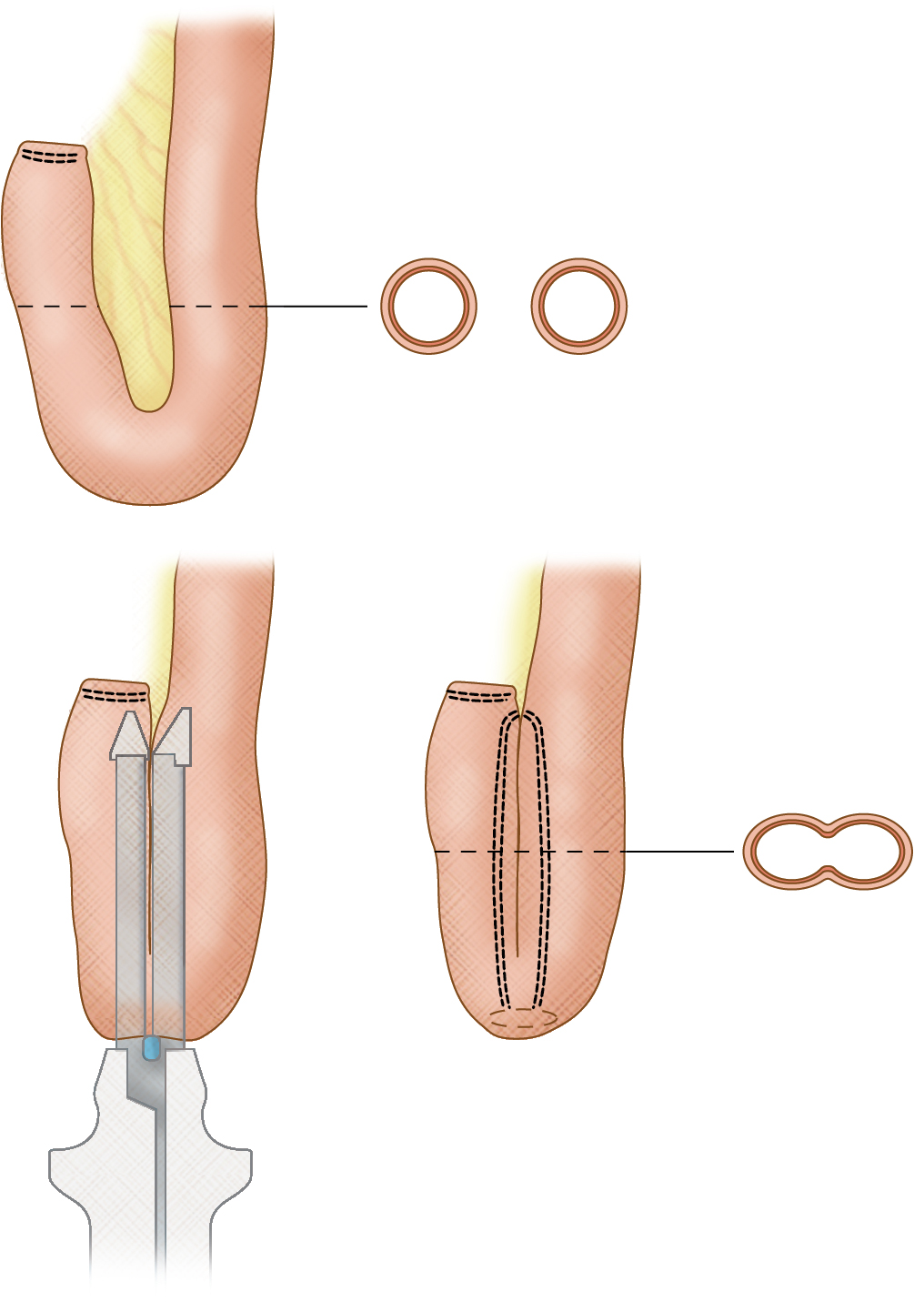
Indications for surgery particular to the colon include complicated diverticulitis, toxic megacolon, severe colitis, and refractory lower GI bleeds. Either part of or the entire colon may need to be resected depending on the etiology ( Fig. 7.6 ), and in the most extreme of cases a total proctocolectomy is performed, which removes the colon and rectum (this can be followed by an ileal J-pouch with anal anastomosis ( Fig. 7.7 ) or an end ileostomy). If only a section of colon is removed and primarily reanastomosed, a temporizing diverting loop ileostomy may also be performed to help the colon heal, and later reversed. If a section of colon needs to be bypassed, an end colostomy can be performed (and later reversed if the obstruction is relieved).
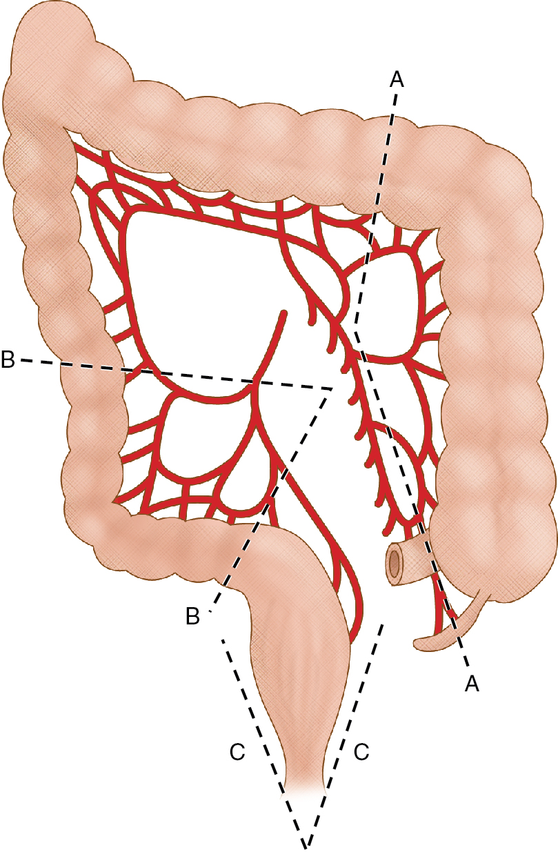
Fig. 7.6
Operative procedures for right-sided colon cancer, sigmoid diverticulitis, and low-lying rectal cancer. Right hemicolectomy involves resection of the terminal ileum and colon up to the division of the middle colic vessels (A). Sigmoidectomy consists of removing colon between the partially retroperitoneal descending colon and the rectum (B). Abdominoperineal resection of the rectum is a combined approach through the abdomen and perineum with resection of the entire rectum (C).
(From Sahani DV, Samir AE. Abdominal Imaging , ed 2. Philadelphia: Elsevier; 2017.)