Catheter
Ultrasound method
Catheter size (F)
Frequency range (MHz)
Depth of field (cm)
Steerability
Doppler/color
ViewFlex PLUSa
Phased-array
9
4.5–8.5
Up to 21
Anterior/posterior, 120°
Yes
AcuNav#
Phased-array
8 or 10
5.0–10
Up to 1
Anterior/posterior
Left/right, 160°
Yes
Phased-Array Catheter
A phased array probe has multiple small elements at the probe tip that emit a beam, which can be pulsed individually at a computer-calculated timing. This allows for a wide volume region to be swept at a high speed. The pulsed elements emit pressure waves which add up to a single wave front traveling at a set angle. The data from multiple beams is combined as the probe is swept through the examined tissue creating a visual image showing a slice through the object. The AcuNav catheter contains a 64-element phased-array transducer which can penetrate up to 15 cm in the heart. Resolution and depth penetration are inversely related. There is four way directional steering in two bidirectional planes (1) anterior-posterior and (2) right-left. These two knobs allow the operator two steer while the third knob locks the catheter position in a desired position. In addition, the catheter itself can be rotated clockwise or counterclockwise.
Main Views for ICE
Venous access can be obtained either internal jugular or femoral approach. The 90–100 cm catheters advanced through an 8F or 10F venous sheath. ICE access site can be either the ipsilateral or contralateral to the working venous access side. If using the ipsilateral approach, a long 30 cm venous sheath is recommended to reduce entanglement with other devices. Others have recommended contralateral access if the patient weighs less than 35 kg. Closure can be a figure of eight or manual pressure. Infusion of 500 cc saline prior to the procedure dilates the veins and chambers; thus assisting with probe advancement and later device placement. The rigid ICE probe without any flexion should be advanced under fluoroscopic guidance as the probe tip is blunt and can puncture or dissect the vein. There are four traditional views (home, septal, long axis, short axis) used during intracardiac interventions (Fig. 13.1). The chamber closest to the beam and at the top of the display screen is always the chamber where the probe is positioned, usually the right atrium.
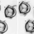
Fig. 13.1
ICE standard views. (a) Schematic diagram of heart. LA left atrium, LV left ventricle, A aorta, PA pulmonary artery. (b, d, f, h) show ICE beam projection using anteroposterior fluoroscopy and corresponding ICE images (c, e, g, i). (b, c) Home view; (d, e) Septal view; (f, g) Long axis view; (h, i) Short axis view
Home View
The home view is obtained by advancing the probe into the mid right atrium without any flexion and the transducer facing the tricuspid valve. Counter clockwise rotation of the catheter may be needed. The right atrium, tricuspid valve, right ventricle, right ventricle outflow tract, aortic valve, anterior part of the atrial septum, pulmonary valve and the proximal part of the pulmonary artery can be visualized in the home view. Tricuspid and pulmonic insufficiencies are best seen in this view. If the catheter is rotated clockwise, the beam is swung posteriorly allowing you to visualize the left atrium, left atrial appendage, mitral valve, pulmonary veins, and proximal descending aorta.
Septal View (Also Called Atrial View)
From the home view, rotate the catheter counterclockwise until the atrial septum is seen. At this point, the catheter is locked and a posterior tilt performed to point the beam cephalad. Both atria are visualized and this is the only view to see the superior rim of the atrial septum. If the beam is moved cephalad with a right tilt, the superior cava is seen on the right side of the screen. The inferior vena cava is seen by moving the probe caudal. Pulmonary venous return to the left atrium and the coronary sinus can be seen in the view.
Long Axis View (Also Caval View)
The long axis view is the locked septal view but the posterior flexed catheter is more cephalad to see the superior vena cava (SVC) or caudal to visualize the Inferior vena cava (IVC). The cephalad position allows better visualization of the SVC, superior aspect of the atrial septum and posterior superior rim. The caudal positioning views the IVC, inferior aspect of the atrial septum and the posterior inferior rim. Clockwise or counterclockwise rotation or posterior-anterior flexion shows right and left pulmonary veins.
Short Axis View
The short axis view is obtained by moving the catheter cephalad below the aortic valve and towards the tricuspid valve. The posterior-anterior knob is adjusted to have less flexion while the left-right knob is rotated more leftward. This allows visualization of the aortic and pulmonary valves and the superior anterior rim (closest to aortic valve) and inferior-posterior rim.
ICE Guidance for Intracardiac Interventions
ASD Evaluation
Figure 13.2 shows the best views to visualize the atrial septals rims for Atrial Septal Defect (ASD) evaluation. The septal view shows the inferior-posterior rim and superior anterior rim. The long axis view allows for visualization of the inferior and superior rims while the long axis view lays out the posterior and anterior rims.
Fig. 13.2
Secundum ASD closure. (a) ICE catheter in septal view position. (b) Septal view. (c) Color Doppler. (d) Rosewire across interatrial septum (e) 30 mm sizing balloon. (f) LAO cran view to measure tunnel length and width. (g) Deployment left atrial side 18 mm Amplatzer ASO. (h) Tug test. (i) Deployed Amplatzer. J. Deployed Amplatzer in LAO cran
Examples Using Intracardiac Echocardiography
Secundum Atrial Septal Defect Closure
The patient shown in Fig. 13.2 was a 74 year old gentleman with tachy-brady syndrome requiring a permanent pacemaker, remote surgical ventricular septal defect repair with NHYA Class III dyspnea. Echocardiography revealed severe bi-atrial enlargement, mitral valve prolapse with moderate mitral regurgitation, and atrial septal defect with predominately left to right shunting. The secundum ASD measured 1.6 cm with the following rims measurements: aortic 26 mm, superior 40 mm, retroaortic 24 mm, posterior 40 mm, inferior vena cava 51 mm and superior vena cava 42 mm.
Related posts:

Stay updated, free articles. Join our Telegram channel

Full access? Get Clinical Tree


