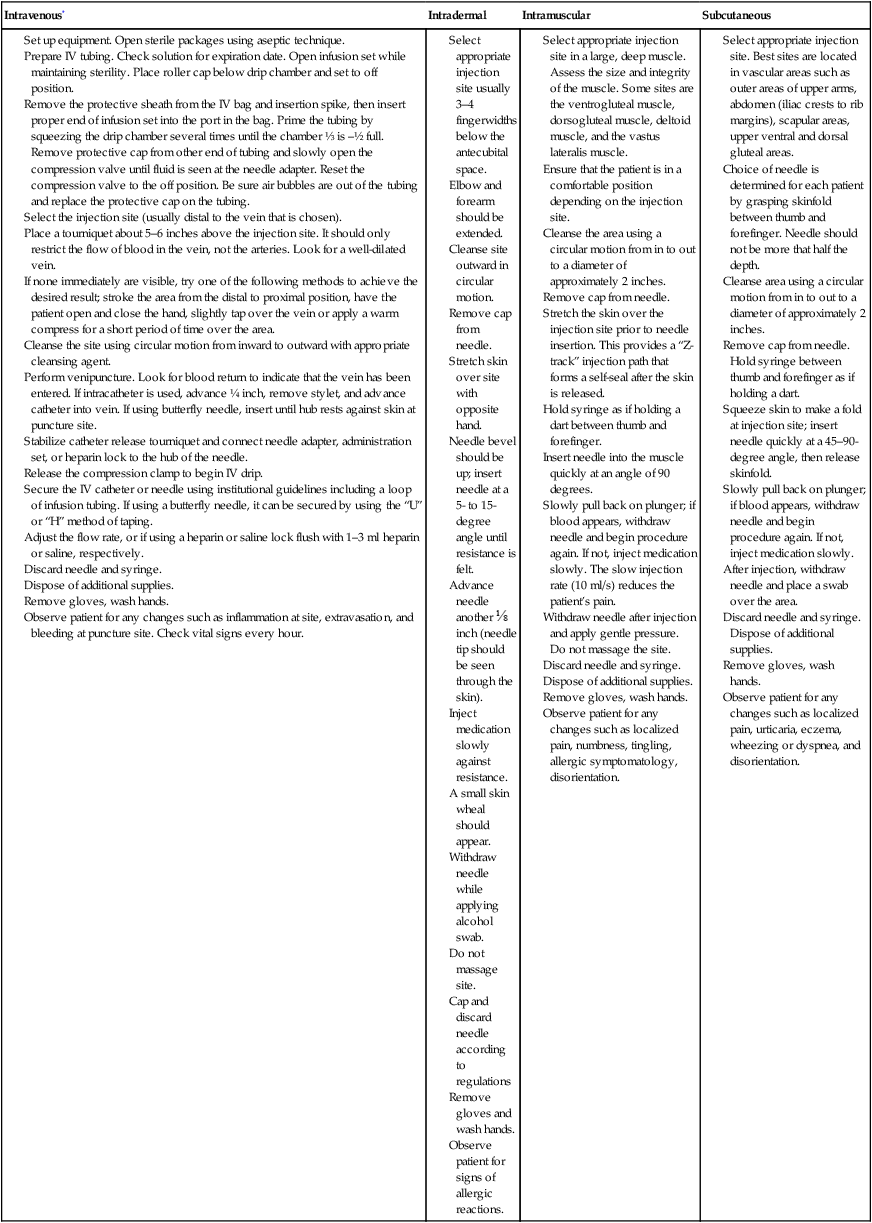
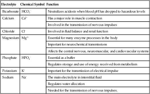
Introduction to Pharmacology

CHAPTER 5


Introduction to Pharmacology


Throughout the 1990s it was sufficient for the special procedure radiographer to have a solid knowledge of the types of contrast agents as well as their action, purpose, and effects with regard to the patient. Beyond this, the broader realm of pharmacology was left to the physician and special procedure nurse. More recently, the practice of diagnostic and interventional radiography requires that the technologists be aware of the types and effects of various medications that are used in the procedures. These include drugs that are prescribed as preexamination medications, those used through the procedure, and the drugs that are available for the treatment of patient reactions. It is beyond the scope of this text to present an in-depth discussion of pharmacology; however, the basic principles of this science will be presented.


The American Registry of Radiologic Technologists (ARRT) requires the candidate for the advanced level examinations to have some understanding regarding the types and administration routes, indications, contraindications, and complications of various drugs used in cardiac and vascular interventional radiography.1 Contrast agents represent a special category of materials; they will be discussed separately in Chapter 6.


TERMINOLOGY


Agonists and Antagonists


When a drug is introduced into the body its action usually begins when it attaches or binds itself to a specific target cell. Four common protein targets are considered binding sites for drugs. These are considered to be regulatory proteins such as enzymes, carrier molecules, ion channels, and receptors. This is a gross simplification of the mechanism of drug action; however, the interaction of the drug and receptor is an important beginning step in understanding the mechanism of drug action and cellular response.


When a drug binds itself to the receptors on specific cells or tissues and produces an effect, it is termed an agonist. On the other hand, the classifications of drugs that bind with certain receptor sites and prevent an action from happening are called antagonists Antagonists are considered “blocking” drugs that bind to the receptor and prevent other drugs from producing an effect. They are also used at times to counteract the action of an agonist. This use or mechanism is called competitive antagonism because each class of drugs is competing for the receptor sites. The overall result will depend upon which class binds to the most receptor sites.


Drug Classification


This refers to the system used to identify drugs. Medications can be classified by their name(s), by their mode of action, or as prescription or nonprescription medications. Each drug that is developed begins its life with a chemical name, the nomenclature used to describe its chemical makeup. Once the chemical has been approved for availability on the commercial market, it is given its generic name. The generic name is derived from the chemical formula in some way and is considered to be nonproprietary. This name indicates that the basic chemical composition of the drug can be provided by several manufacturers. When a pharmaceutical house trademarks a name for its specific brand of the drug, it can then be classified by its trade or brand name.


Pharmacology is the study of drugs and their origins, chemical composition, preparations, and use. Pharmacokinetics refers to the mechanisms of bodily absorption, distribution, metabolism, and excretion of drugs. Pharmacodynamics is the action that various drugs have with body tissues.


ROUTES OF ADMINISTRATION


The route of administration will vary not only with the drug used but also the purpose that it has been designed for. The drugs used in vascular and cardiac diagnostic and interventional radiography fall into two major classifications: local and systemic.


The local medications are usually administered at a specific site and are injected into the tissues only in that particular area. The route is by direct injection, and the purpose of the drug is the reduction of sensation (pain) in the tissues of the surrounding area. These drugs are used at the beginning of the procedure, and they have an anesthetic and analgesic effect at the puncture site. The anesthetic effect of the “local” medication is almost immediate and is well tolerated by most adult patients. Box 5-1 lists some local anesthetics. Occasionally, one of the amides will be added to the contrast agent to reduce the discomfort and pain associated with the injection.


Local medications can also be administered by inhalation and topical application to the skin and other mucous membranes. Inhaled medication is used to produce a rapid response to local respiratory conditions. The glucocorticoids and bronchodilators are administered by inhalation. Despite the fact that these drugs are given for local conditions, some of the medication will enter the systemic circulation and can be the cause of various side effects. Topical administration is a painless way of administering some medications with the probability of limited consequence.


Systemic medications produce a wide variety of effects to the patient. These drugs are usually used before the procedure begins, at times during the procedure, and often in emergency situations to alleviate a problem. The four major routes of administration for the systemic agents are oral, rectal, sublingual, and parenteral. The first three routes are self-explanatory, and their use will appear obvious. Inhalation can also be considered as a means of administering systemic medications as in the case of general anesthesia.


Oral medications are taken by mouth. The rectal route of administration is used if the patients are unable to take oral medication. This could be due to the inability to retain the drug in the stomach or because the drug would be adversely affected by the gastric contents. Sublingual administration of certain medications is used when quick action of the drug is required. These drugs are manufactured to dissolve quickly when placed under the tongue to provide a rapid effect. It can be seen that these three routes of administration of medication all utilize the digestive system to introduce the medication into the body. The parenteral route pertains to the administration of a medication other than the gastrointestinal tract. This is usually accomplished by injection.


There are five subdivisions of parenteral administration of medication: intravenous, intramuscular, intrathecal, intradermal, and subcutaneous. Each of these methods requires the use of a needle and usually a syringe. Intravenous (IV) fluid therapy is the exception to the use of a syringe as a part of the process. Refer to Chapter 4 for the discussion regarding needles and syringes.


Each of the parenteral routes of administration is similar in that the object of the procedure is to place a needle into a specific part of the body. Table 5-1 summarizes the locations for the injections as well as any specifics about the process.


General Guidelines for Parenteral Medication Administration


In this section we will discuss the general guidelines for the administration of parenteral medication. Table 5-1 illustrates the major differences in the various parenteral routes of medication administration. As mentioned earlier, medications can be administered to the patient in a variety of different ways: oral, rectal, topical, inhalation, and parenteral. The parenteral route of drug administration is subdivided into five separate areas: intravenous, intradermal, intrathecal, subcutaneous, and intramuscular.


The guidelines will be essentially the same for all routes of administration with the exception that the parenteral administration of medications is considered an invasive procedure and all standard precautions should be observed during the process. Strict aseptic techniques should be employed. The specifics of administration for each of the routes should be meticulously followed. The radiographer not only must be aware of the specifics of each parenteral route but must guard against inadvertent personal needle stick injury.


When any medication is administered it is important to follow some basic principles, known as the “Six Rights.”2 These basic rules should be followed whenever any medication is dispensed to a patient.


As a part of the advanced procedure team, it is imperative to have a working knowledge of all of the medications commonly used for the specific procedure. This also includes any emergency drugs. In other words the radiographer must be knowledgeable about the medications given to the patient before, during, and after the procedure. This practice will help ensure that the patient will have the greatest possibility for a successful and uneventful procedure.


Many of the guidelines listed here are the same for routine patient care and communication. The difference is that all of the vascular and cardiac interventional procedures are invasive and have the potential to produce a number of different reactions. Extreme care should be exercised in adhering to these rules and guides. In almost every case some type of medication is administered to the patient before, during, or after a procedure. It is the responsibility of everyone on the team to practice a heightened level of care in order to ensure patient safety from medication reactions due to human error. The Institute for Safe Medication Practices (ISMP) has identified some common medications that have been known to cause an increased risk for patient injury when errors are made. The organization recommends that when any of these drugs are administered safety strategies are in place to prevent any errors. Box 5-2 summarizes the ISMP List of High Alert Medications.3


BOX 5-2   ISMP List of High Alert Medications


Class/Category of Medications


image Adrenergic agonists, IV (e.g., epinephrine)


image Adrenergic antagonists, IV (e.g., propranolol)


image Anesthetic agents, general, inhaled, and IV (e.g., propofol)


image Cardioplegic solutions


image Chemotherapeutic agents, parenteral and oral


image Dextrose, hypertonic, 20% or greater


image Dialysis solutions, peritoneal and hemodialysis


image Epidural or intrathecal medications


image Glycoprotein IIb/IIIa inhibitors (e.g., eptifibatide)


image Hypoglycemics, oral


image Inotropic medications, IV (e.g., digoxin, milrinone)


image Liposomal forms of drugs (e.g., liposomal amphotericin B)


image Moderate sedation agents, IV (e.g., midazolam)


image Moderate sedation agents, oral, for children (e.g., chloral hydrate)


image Narcotics/opiates, IV and oral (including liquid concentrates, immediate and sustained release)


image Neuromuscular blocking agents (e.g., succinylcholine)


image Radiocontrast agents, IV


image Thrombolytics/fibrinolytics, IV (e.g., tenecteplase)


image Total parenteral nutrition solutions


Institute for Safe Medication Practices: ISMP’s List of High Alert Medications, 2003, http://www.ismp.org/MSArticles/highalert.htm


Parenteral administration (by injection and not via the gastrointestinal tract) requires additional guidelines depending upon the subclassification of the injection route. The first 10 steps in any procedure for the administration of medication would be the same and are as follows:


Table 5-2 lists the procedures for each of the parenteral routes of administration with the exception of the intrathecal route, which is discussed in detail in Chapter 21.


TABLE 5-2


Generalized Guidelines for Parenteral Administration of Medication


Intravenous* Intradermal Intramuscular Subcutaneous


Set up equipment. Open sterile packages using aseptic technique.


Prepare IV tubing. Check solution for expiration date. Open infusion set while maintaining sterility. Place roller cap below drip chamber and set to off position.


Remove the protective sheath from the IV bag and insertion spike, then insert proper end of infusion set into the port in the bag. Prime the tubing by squeezing the drip chamber several times until the chamber ⅓ is –½ full. Remove protective cap from other end of tubing and slowly open the compression valve until fluid is seen at the needle adapter. Reset the compression valve to the off position. Be sure air bubbles are out of the tubing and replace the protective cap on the tubing.


Select the injection site (usually distal to the vein that is chosen).


Place a tourniquet about 5–6 inches above the injection site. It should only restrict the flow of blood in the vein, not the arteries. Look for a well-dilated vein.


If none immediately are visible, try one of the following methods to achieve the desired result; stroke the area from the distal to proximal position, have the patient open and close the hand, slightly tap over the vein or apply a warm compress for a short period of time over the area.


Cleanse the site using circular motion from inward to outward with appropriate cleansing agent.


Perform venipuncture. Look for blood return to indicate that the vein has been entered. If intracatheter is used, advance ¼ inch, remove stylet, and advance catheter into vein. If using butterfly needle, insert until hub rests against skin at puncture site.


Stabilize catheter release tourniquet and connect needle adapter, administration set, or heparin lock to the hub of the needle.


Release the compression clamp to begin IV drip.


Secure the IV catheter or needle using institutional guidelines including a loop of infusion tubing. If using a butterfly needle, it can be secured by using the “U” or “H” method of taping.


Adjust the flow rate, or if using a heparin or saline lock flush with 1–3 ml heparin or saline, respectively.


Discard needle and syringe.


Dispose of additional supplies.


Remove gloves, wash hands.


Observe patient for any changes such as inflammation at site, extravasation, and bleeding at puncture site. Check vital signs every hour.


Select appropriate injection site in a large, deep muscle. Assess the size and integrity of the muscle. Some sites are the ventrogluteal muscle, dorsogluteal muscle, deltoid muscle, and the vastus lateralis muscle.


Ensure that the patient is in a comfortable position depending on the injection site.


Cleanse the area using a circular motion from in to out to a diameter of approximately 2 inches.


Remove cap from needle.


Stretch the skin over the injection site prior to needle insertion. This provides a “Z-track” injection path that forms a self-seal after the skin is released.


Hold syringe as if holding a dart between thumb and forefinger.


Insert needle into the muscle quickly at an angle of 90 degrees.


Slowly pull back on plunger; if blood appears, withdraw needle and begin procedure again. If not, inject medication slowly. The slow injection rate (10 ml/s) reduces the patient’s pain.


Withdraw needle after injection and apply gentle pressure. Do not massage the site.


Discard needle and syringe.


Dispose of additional supplies.


Remove gloves, wash hands.


Observe patient for any changes such as localized pain, numbness, tingling, allergic symptomatology, disorientation.


Select appropriate injection site. Best sites are located in vascular areas such as outer areas of upper arms, abdomen (iliac crests to rib margins), scapular areas, upper ventral and dorsal gluteal areas.


Choice of needle is determined for each patient by grasping skinfold between thumb and forefinger. Needle should not be more that half the depth.


Cleanse area using a circular motion from in to out to a diameter of approximately 2 inches.


Remove cap from needle. Hold syringe between thumb and forefinger as if holding a dart.


Squeeze skin to make a fold at injection site; insert needle quickly at a 45–90-degree angle, then release skinfold.


Slowly pull back on plunger; if blood appears, withdraw needle and begin procedure again. If not, inject medication slowly.


After injection, withdraw needle and place a swab over the area.


Discard needle and syringe. Dispose of additional supplies.


Remove gloves, wash hands.


Observe patient for any changes such as localized pain, urticaria, eczema, wheezing or dyspnea, and disorientation.


image


*A discussion of venipuncture and the use of heparin or normal saline locks is presented in the section, “Intravenous Therapy.”


Intravenous Therapy


Intravenous therapy involves the introduction of a fluid through a vein. This is accomplished using an intravenous cannula such as a needle or one of the special angiocatheters. The location or insertion site can be either in a central or a peripheral vein. In most cases the peripheral sites are used. These include the veins of the hand; the veins of the arm (antecubital, radial, ulnar, cephalic), and the external jugular vein.


The main purpose of this type of therapy is the management of the fluids in the body. The body is composed of over 65% water, and fluid management is vitally important for the balance of electrolytes, blood volume, and other nutritional materials required for intra- and extracellular equilibrium. In the normal, healthy patient this is accomplished automatically even under situations in which there is a minor fluid loss. Extracellular fluids are the body fluids consisting of the interstitial fluid and blood plasma. Approximately one quarter of the body’s water is located outside the cells. Most of this water is found in the interstitial space. Interstitial fluid fills the spaces between most of the cells of the body. Less than 8% of the body’s fluid is located in the intravascular space. Three quarters of all the water is found within the cells of the body. These intracellular fluids are within cell membranes throughout the body and contain dissolved solutes essential to fluid and electrolyte balance and metabolism. Electrolytes are substances that ionize when dissolved in water and as such are able to conduct a current. Calcium is an electrolyte that is necessary for the conduction of nerve impulses responsible for the contraction of skeletal muscle. All of these substances are necessary for normal body metabolism. Table 5-3 lists the various electrolytes present in the body’s fluids.


The therapeutic introduction of fluid materials becomes necessary under certain conditions, such as trauma, surgical procedures, illness, and factors contributing to an increased level of stress. Ernest H. Starling, a British physiologist, proposed that a balance exists at the level of the capillary membranes when the fluid entering the circulation and that leaving the circulation are the same. His theory described the capillary wall as a semipermeable membrane that allowed salt solutions to pass through it (by osmosis). In order to balance the concentration on both sides of the membrane, the osmotic pressure forced movement of fluids from the tissue into the circulation. In other words, the fluids will move toward the side of the semipermeable membrane that contains the largest proportion of these solutes.


Osmolarity and Osmolality


Certain concepts must be understood prior to a discussion of intravenous therapy. Starling’s principle is not only applicable to intravenous therapy; it is thought that many of the reactions caused by the administration of contrast media are the result of this osmotic action. The terms osmolarity and osmolality are used quite extensively in describing different solutions, including the various classes of positive contrast media, and must be understood in order to understand their actions within the body. A review of osmosis is necessary in order to understand the concepts of osmolarity and osmolality.


Osmosis is defined as the passage of a solvent (water) through a semipermeable membrane into a solution of higher solute (a salt or a substance dissolved in a solution) concentration that tends to equalize the concentrations of solute on the two sides of the membrane. In the body, human plasma is considered to be isotonic. In order to maintain this isotonic state, everything administered into the body must be equated to the sodium chloride equivalent of the human plasma, because sodium chloride is a major determinate of blood osmolarity.


Any substance dissolved in a solution can be considered a solute. Although osmosis is not limited to the actions resulting from the administration of IV therapy solutions or contrast media, the discussion of this concept will relate to the action of water as the solvent and contrast medium as the solute. Osmosis is the tendency for a solvent, such as water, to move through a permeable or semipermeable membrane so as to become mixed with a solute, such as contrast medium, to create a solution that is equal in osmotic pressure on both sides of the membrane. This movement of the solvent (water) is from a solution that is lower in solute (contrast medium) concentration to the side of the membrane that is higher in solute (contrast medium) concentration. Osmotic activity will continue until the concentration of the solutions on both sides of the membrane is equalized. In essence, osmosis is the movement of water through the membranes separating the body’s compartments.


Osmolarity and osmolality refer to the osmotic pressure of a solution. This pressure is the mechanism that controls the movement of the water across the membrane. Osmolarity refers to the concentration per volume of solution (milliosmols per kilogram of solution), while osmolality refers to weight or milliosmols per kilogram of water. These two terms are often used interchangeably, and although they both relate to osmotic pressure it can be seen that they are actually different measurements. In most cases, there is little difference in these measurements; however, in certain cases the discrepancy can be considerable. Osmolality is the term that should be applied when discussing the intravenous therapy solutions and radiopaque contrast media.


An important fact is that the solute does not usually pass through the membrane and only the solvent moves to equalize the solution’s concentration. Under certain circumstances, however, the solute can filter into the interstitial fluid via endothelial cell clefts. This can occur because of the high osmolality of the mixture of the solute and blood.


In the central nervous system the endothelium has a continuous basement membrane, and the cells are connected by tight junctions. This is referred to as the blood-brain barrier. The blood-brain barrier will normally hold back the passage of a number of solutes serving as a protective guard to the body. The blood-brain barrier is the anatomic/physiologic aspect of the brain that separates the parenchyma (organ tissue) of the central nervous system from the blood. Current theory is that the blood-brain barrier prevents or slows the passage of various chemical compounds and other potentially harmful substances from the blood into the tissue of the central nervous system.


The ultimate goal of intravenous therapy is to prevent or correct fluid and electrolyte (solute) disturbances. Depending upon the purpose, intravenous fluids can be hypotonic, hypertonic, or isotonic. Solutions that are similar in content to physiologic fluids are said to be isotonic. Depending upon the source of information, an isotonic solution ranges between 240 and 375 mOsm. Isotonic solutions are also considered to be equivalent to a 0.9% weight/volume NaCl solution, which is equivalent to that of blood plasma. This type of solution does not exert any osmotic action in either direction at the capillary level.


Hypertonic solutions generally are greater than 375 mOsm. In the attempt to achieve balance, these solutions will move fluid across the semipermeable membrane at the capillary level and into the intravascular spaces, causing the cells to shrink and get smaller. These solutions have a greater solute concentration than blood plasma, and fluid is needed to achieve balance.


When a solution is less than 240 mOsm it has lower concentration of solutes than blood plasma and is considered hypotonic. In order to achieve balance, the fluid from the vascular space moves across the semipermeable membrane and into the cells. This influx has a tendency to cause the cells to expand and get larger.


The importance of the definitions listed here is that the technologist should be aware of the osmolality of the solution that is being introduced into the patient. The IV solution packs, referred to as “bags,” are labeled with the osmolality. This can be easily compared with the range for isotonic solutions, and the radiographer can quickly determine which of the three types of solutions is being administered.


Intravenous therapy when used during advanced radiologic procedures serves several functions. Among these functions, the intravenous port provides a ready access to the vein for the rapid introduction of emergency medication; however, fluid management is the primary purpose, especially when contrast agents are administered. The patient should be well hydrated during contrast examinations to reduce the probability of an adverse reaction. The patient’s condition and the type and amount of contrast agent will govern the type and flow rate of the intravenous fluid. Box 5-3 summarizes the reasons that IV therapy is employed.


Venipuncture.

The general guidelines for initiating an IV line have already been discussed for intravenous fluid replacement utilizing a metal needle and extension tubing for connection to a bag or bottle of IV fluid. Depending upon the nature or purpose for entering the vein the difference between the collection of a specimen or the introduction of a bolus of medication via a vein, and placing a long-term access port is minimal.


Venipuncture is generally accomplished utilizing a metal needle attached to a syringe or a device called a Vacutainer tube. In either case the process is the same. The steps are as follows:


Feb 27, 2016 | Posted by in GENERAL RADIOLOGY | Comments Off on Introduction to Pharmacology

Full access? Get Clinical Tree

Get Clinical Tree app for offline access