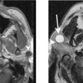
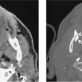
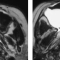
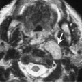
Chapter 11 Malignant fibrous histiocytoma was uncommonly reported before the 1970s. Since then, this tumor has become the most frequently reported soft tissue sarcoma in most major institutions. About 3 to 10% of all malignant fibrous histiocytoma occur in the head and neck. There is no sex predilection and the tumor occurs most frequently in adults 50 to 60 years of age. Patients usually present with a large mass, with or without pain. Symptoms and signs depend on the location of the tumor and compression of surrounding structures. These tumors arise from tissue histiocytes. They grow and invade along fascial planes. They usually have well-defined margins but are not encapsulated. The verifying histologic picture is the storiform pattern of fibroblastic and histiocytic cells. Most tumors show cellular pleomorphism with multiple giant cells. The prognosis is poor. There is a very high recurrence rate following surgery. The tumor is radioresistant and does not respond significantly to chemotherapy. Tumors also show a high propensity to metastasize. Malignant fibrous histiocytoma appears as a well-defined soft tissue mass with moderate contrast enhancement (Figs. 11–1 and 11–2). It may cause erosion of adjacent bony structures and may spread intracranially via neural foramina.
Malignant Fibrous Histiocytoma
Epidemiology
Clinical Findings
Pathology
Treatment
Imaging Findings
CT
Related posts:

Stay updated, free articles. Join our Telegram channel

Full access? Get Clinical Tree


