NASOPHARYNX: INFECTIONS
KEY POINTS
- Imaging, especially computed tomography, may be useful in the management of selected nasopharyngeal infections.
- Computed tomography can provide a safe and rapid assessment of the airway in a patient who is stable and under the control of the treating physician for airway management decisions.
- Atypical clinical and imaging features of nasopharyngeal pathology should raise the possibility of an inflammatory process.
Most acute infections of the pharynx present an obvious clinical picture, and imaging is reserved for establishing the extent rather than the cause of the disease, such as whether pyogenic adenoiditis is associated with oropharyngeal infection and a tonsillar or peritonsillar abscess. The very important exception to this is suppurative retropharyngeal adenitis, which is discussed in detail in Chapter 148. That condition should not be mistaken for a retropharyngeal abscess since it is almost completely responsive to medical therapy without any need for surgical intervention.1
After physical examination and in less definitive clinical situations, it may be difficult to decide whether an area of nasopharyngeal swelling is inflammatory or infectious as opposed to neoplastic under less clear clinical circumstances. For instance, in a low-grade inflammatory process, this distinction might remain unclear even after biopsy and watchful waiting might become the default strategy, sometimes with imaging surveillance as an aid.
The nasopharynx may be the primary source of skull base osteomyelitis (Chapters 114 and 115) or secondarily involved by that disease. Imaging will be the prime determinant of the nature and extent of such usually chronic infections and may be used as guidance for tissue sampling and the definitive diagnosis.
Much inflammatory swelling in the nasopharynx will have a nonspecific imaging appearance when considered independent of the clinical setting. Taken with the clinical setting, the findings are often specific enough to confirm the etiology suspected clinically. This chapter considers infections, while noninfectious inflammatory conditions are presented in Chapter 187.
ANATOMIC AND DEVELOPMENTAL CONSIDERATIONS
Embryology
Occasionally, a developmental lesion such as a branchial apparatus cyst (Chapter 153) will become infected and present as a primary nasopharyngeal infection (Figs. 185.1F and 185.2A,B and Chapter 185).
Applied Anatomy
A thorough knowledge of nasopharyngeal anatomy and anatomic variations is required for the evaluation of infections of the nasopharynx. This anatomy is presented in detail with the introductory material on the nasopharynx (Chapter 184). These conditions are often contiguous with the skull base and temporal bone (Chapter 104); oropharynx (Chapter 190); and retropharyngeal, parapharyngeal, and masticator spaces (Section VI on the suprahyoid neck). Those chapters should be reviewed for both anatomic highlights and spread patterns of disease to develop a complete understanding of the regional spread patterns and related complications of these infections.
IMAGING APPROACH
Techniques and Relevant Aspects
Computed Tomography and Magnetic Resonance Imaging
Inflammatory conditions of the nasopharynx are studied with computed tomography (CT) and magnetic resonance imaging (MRI) in essentially the same manner as benign and malignant nasopharyngeal tumors, except the entire neck may not be included. The specifics and relative value of using these studies in this anatomic region are reviewed in Chapter 185. Problem-driven protocols for CT and MRI are presented in Appendixes A and B.
Other
Ultrasound has no definitive role in the evaluation of these infections, although intraoral ultrasound has been reported as useful for detecting possible peritonsillar abscess and suppurative retropharyngeal nodes.2 Radionuclide studies to evaluate infectious disease (Chapter 5) have modest diagnostic use in the evaluation of nasopharyngeal conditions when those infections are associated with skull base osteomyelitis (Chapters 114 and 115). In those infections, their main utility is to monitor the therapeutic response of a chronic infection.
Plain film soft tissue views of the neck, while often done, are typically not definitive and delay the diagnostic process by sometimes leading to a false sense of security.
Pros and Cons
Infections in this area may involve or arise in the skull base so that CT becomes immediately preferable to MR. The patient with an acute infection may be very uncomfortable and MRI motion degraded. A definitive CT study providing all necessary information can be accomplished in about 5–10 minutes making it a more sensible first choice. MRI can be used for problem solving such as in chronic conditions where it might help choose the most productive tissue or fluid sampling site when it is difficult to differentiate tumor from a chronic inflammatory process.
SPECIFIC DISEASE/CONDITION
Acute and Subacute Pyogenic Bacterial Infections, Viral Infections, and Chronic Infections
Etiology
The vast majority of infections of the nasopharynx that come to imaging are acute or subacute pyogenic bacterial infections producing suppurative retropharyngeal adenitis1 (Fig. 186.1 and Chapter 148). The other group in this category is upper pharyngeal infections that might be complicated by abscess formation (Chapter 192).
Fungal infection occurs predominantly in the diabetic and otherwise immune-compromised population (Fig. 186.2). Tuberculosis (TB) may involve the Waldeyer ring.
Viral infections are usually not so severe that they clinically mimic pyogenic infection enough to trigger imaging with CT or MRI (Fig. 186.3). Epstein-Barr virus infection, including posttransplant lymphoproliferative disease, may mimic lymphoma, and the related adenopathy may trigger imaging. Human immunodeficiency virus (HIV) infection can manifest in the lymphoid tissue of the Waldeyer ring (Figs. 186.4 and 186.5).
Prevalence and Epidemiology
Pyogenic bacterial infections occur sporadically, primarily in the pediatric and young adult populations. In the older age group diabetes, especially when poorly controlled, is a predisposing factor to what might be very aggressive pharyngeal infections (Fig. 186.6). HIV and other immune-compromising conditions will predispose to fungal infections and TB (Fig. 186.2). Infections may also be a complication of surgery such as adenoidectomy (Fig. 186.7).
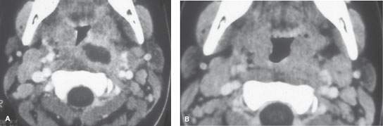
FIGURE 186.1. Contrast-enhanced computed tomography (CT) study of a patient with suppurative adenitis due to pharyngitis. Pretreatment CT is seen in (A). The patient was treated with intravenous antibiotics, and this follow-up study (B) was obtained at 72 hours. It shows resolution of the suppurative lymph node in response to antibiotic therapy.
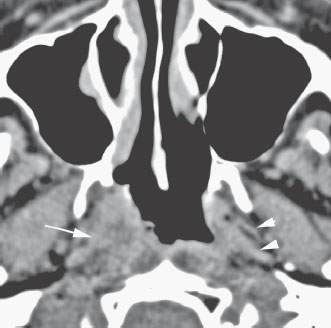
FIGURE 186.2. An immune-compromised patient with a computed tomography study showing an infiltrative process along the right lateral nasopharyngeal wall with obliteration of deep tissue planes (arrow) as compared to the opposite side (arrowheads). This was due to invasive aspergillosis.
Clinical Presentation
In infants, acute and subacute bacterial infections will present with fever and poor feeding and possibly lymphadenopathy. In older children and adults, these infections will cause fever, usually a very intense sore throat and altered voice, and lymphadenopathy. In the more indolent infections and in immune-compromised patients, there may be little, if any, fever, and local symptoms such as odynophagia may be less prevalent.
Pathophysiology and Patterns of Disease
The morphology of infectious and inflammatory disease and related bone and vascular complications are discussed in detail in Chapters 13 through 16.
Manifestations and Findings
Plain Film and Fluoroscopy
Plain films may show pharyngeal swelling, especially along the posterior pharyngeal wall, or extraluminal gas. In general, this is a superfluous first step in a patient that requires a definitive imaging evaluation.
Computed Tomography and Magnetic Resonance Imaging
The details of how these infections spread and tend to generally appear on contrast-enhanced CT are discussed in Chapters 13 through 16.
Contrast-enhanced CT and contrast-enhanced magnetic resonance (MR) will show a pyogenic process to be one that has a typical cellulitis pattern and may show an associated contained or spreading abscess (Chapter 13). Both modalities should almost always be able to differentiate “drainable” from “nondrainable” infectious processes. These findings may spread into the deep planes around the nasopharynx and adjacent regions. Bone erosion (Chapter 14) and vascular complications (Chapter 15) may be present. The most well-known vascular complication is Lemierre syndrome, which most typically is associated with acute pyogenic tonsillitis (Fig. 186.8). Carotid arteritis may be present, but it will not typically be symptomatic. Cortical bone erosion may not be as evident on MR as on CT. More indolent infections may show a similar morphology but are less likely to show frank abscess cavities (Chapter 16). Most of these infections will enhance more than muscle. They replace fat and may spread along paths of least resistance. It is important to understand differentiating features of the following conditions in this category.
In patients with any pyogenic pharyngeal infection that comes to imaging, it is important to search for evidence of internal jugular vein thrombosis; septic thrombosis may lead to metastatic infection, most commonly in the lungs but also in the joints and liver; rarely, the meninges may be involved or other intracranial complications occur. This complication of a primary oropharyngeal infection is known as Lemierre syndrome. These patients have positive blood cultures for Fusobacterium necrophorum, the causative bacterium. If not properly treated, the course of this syndrome may be fatal.3,4
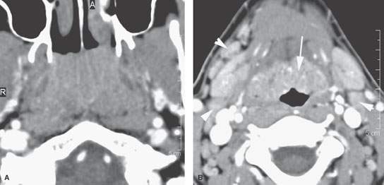
FIGURE 186.3. A patient with viral upper respiratory infection due to Epstein-Barr virus. The inflamed hypertrophied adenoidal tissue fills the nasopharyngeal airway in (A). In (B), inflamed lingual tonsillar tissue (arrow) and reactive lymph nodes are present (arrowheads).
Related posts:
 Preseptal Compartment: Acute and Chronic Infections and Noninfectious Inflammatory Conditions
 Necrotizing (“Malignant”) Otitis Externa
 Cervicothoracic Junction: Brachial Plexus Trauma
 Tumors of the Submandibular Gland and Space and Tumorlike Conditions
 Masticator Space, Buccal Space, and Infratemporal Fossa Infections and Other Inflammatory Conditions
 Oral Cavity and Floor of the Mouth: Malignant Tumors
Stay updated, free articles. Join our Telegram channel

Full access? Get Clinical Tree


