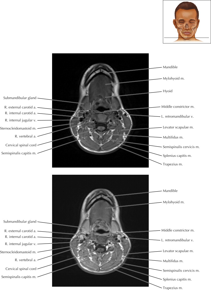
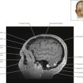
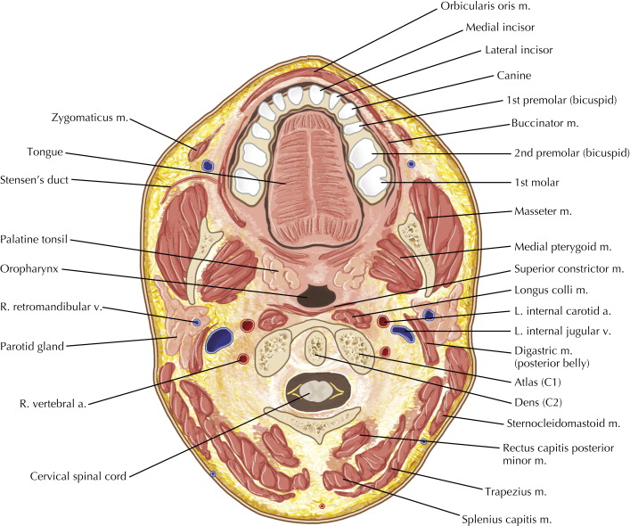
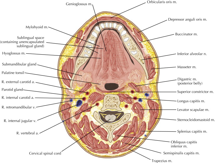
Oral Cavity, Pharynx, and Suprahyoid Neck Axial 1
Normal Anatomy
Note the enhancement of the mucosal surfaces of the aerodigestive tract on the lower magnetic resonance (MR) image in Axial 1 after gadolinium contrast administration. The subcutaneous adipose tissue and the adipose planes between the structures in the neck are visible on both the upper pre-contrast T1-weighted MR image and the lower post-contrast T1-weighted MR image.
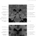
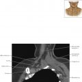
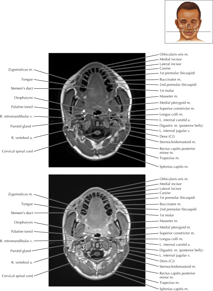
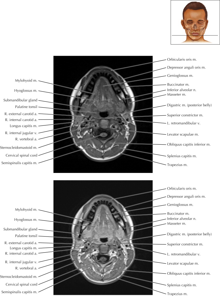
Oral Cavity, Pharynx, and Suprahyoid Neck Axial 2
Pathologic Process
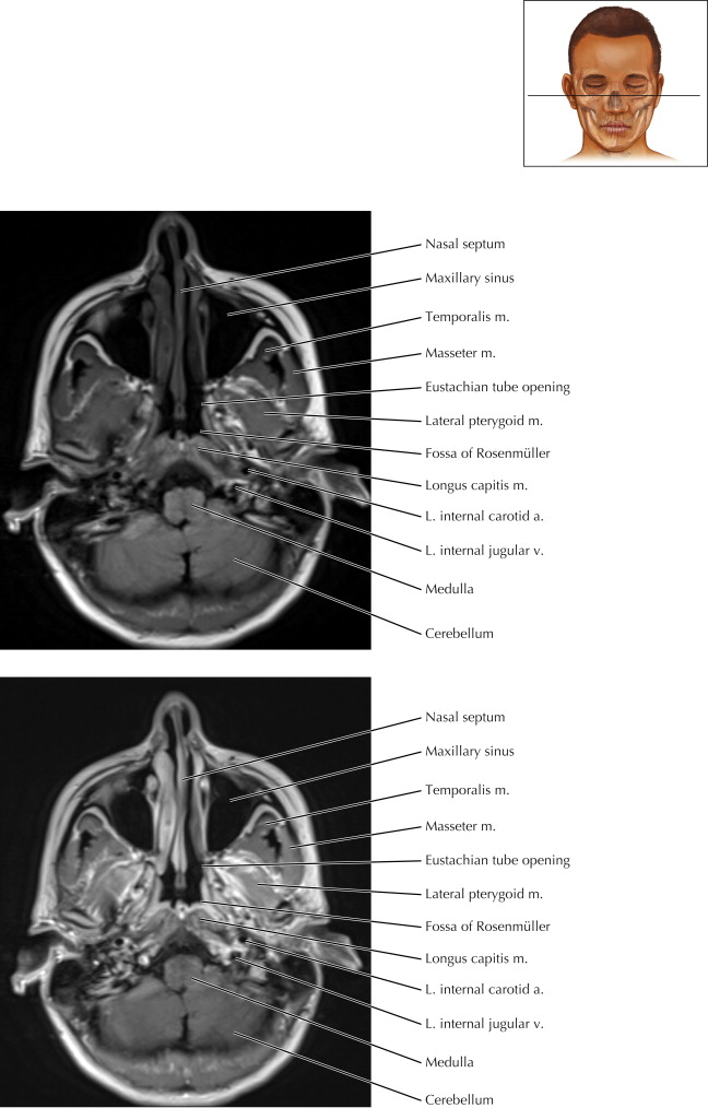
An axial image of the nasopharynx at the level of the foramen magnum is visible on typically every brain study. Fluid in the mastoid air cells on one side may indicate a mass obstructing the Eustachian canal opening anterior to the torus tubarius (posterior protuberance of pharyngeal opening of auditory tube). The fossa of Rosenmüller is a blind-ending divot between the torus tubarius and the longus capitis muscle more posteriorly. Asymmetric loss of this fossa may be the first sign of a nasopharyngeal lesion. Malignant spread of a nasopharyngeal carcinoma may first reach the retropharyngeal lymph node. This lymph node is normally less than 8 mm in long axis and located between the longus capitis muscle and the internal carotid artery more posterolaterally.

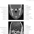
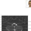
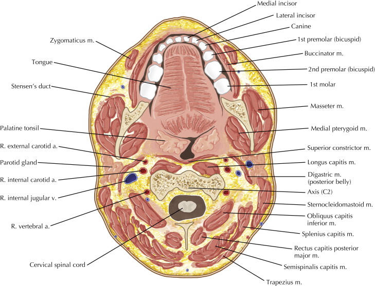
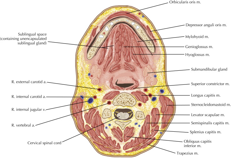
Oral Cavity, Pharynx, and Suprahyoid Neck Axial 5
Pathologic Process
Lesions of the head and neck are often most easily detected by loss of normal fat planes. On these T1-weighted precontrast (upper) and postcontrast (lower) MR images, note the fat within the retromolar trigone (retromandibular triangle) located posterior to the third molar. Once a malignancy or infection has reached the parapharyngeal fat located more posteriorly, it has an easy route of spread craniocaudally from the skull base down to the inferior pericardial recess.
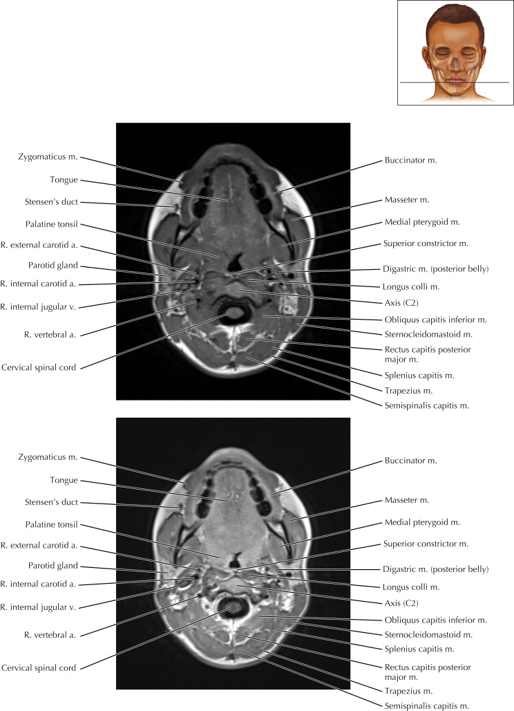
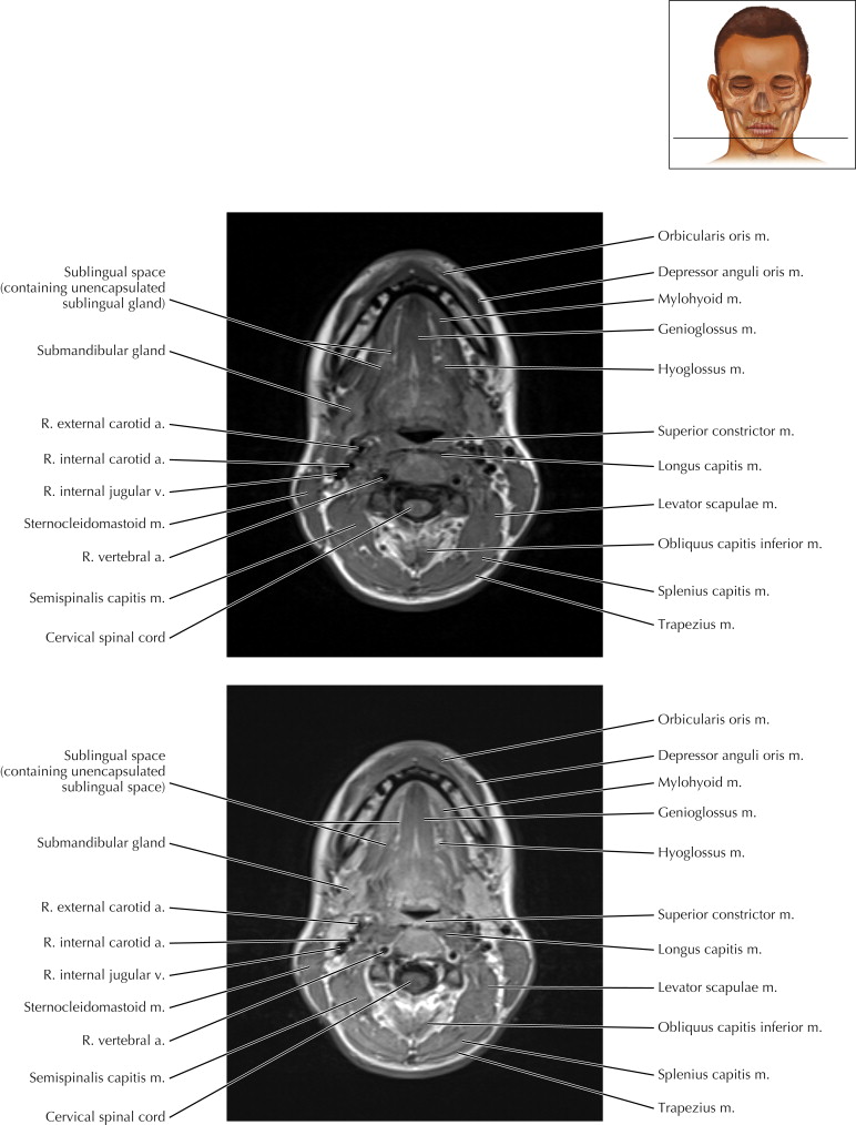
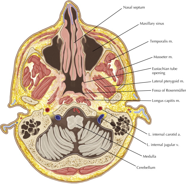
Oral Cavity, Pharynx, and Suprahyoid Neck Axial 8