Frontal bone
Sphenoid bone (lesser wing)
Sphenoid bone (greater wing)
Zygomatic bone
Zygomatic arch
Maxillary bone
Supraorbital foramen
Superior orbital fissure
Optic canal
Nasal bone
Lacrimal bone
Ethmoid bone
Palatine bone
Inferior orbital fissure
Infraorbital foramen
Frontal graphic demonstrates the complex anatomy of the bony orbit. The walls of the orbital cavity receive contributions from 8 different bones of the skull. The complex foramina and fissures at the apex are located primarily within the greater & lesser wings of the sphenoid bone and its junctions with adjacent bones.
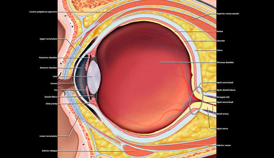
Upper tarsal plate
Posterior chamber
Anterior chamber
Lens
Cornea
Iris
Zonule fibers
Ciliary body
Lower tarsal plate
Inferior oblique
Superior rectus muscle
Retina
Choroid
Sclera
Vitreous chamber
Optic nerve head
Optic sheath (dura)
Perioptic CSF
Optic nerve head
Renal artery
Optic nerve
Inferior rectus
Sagittal graphic demonstrates the anterior and posterior segments of the globe. The aqueous anterior segment is comprised of the anterior chamber and very small posterior chamber. The much larger posterior segment is filled by the vitreous chamber. The layered tunicae of the retina, choroid, and sclera are demonstrated as well as the components of the optic nerve at its insertion. Some of the extraocular muscles and eyelid structures are also demonstrated.
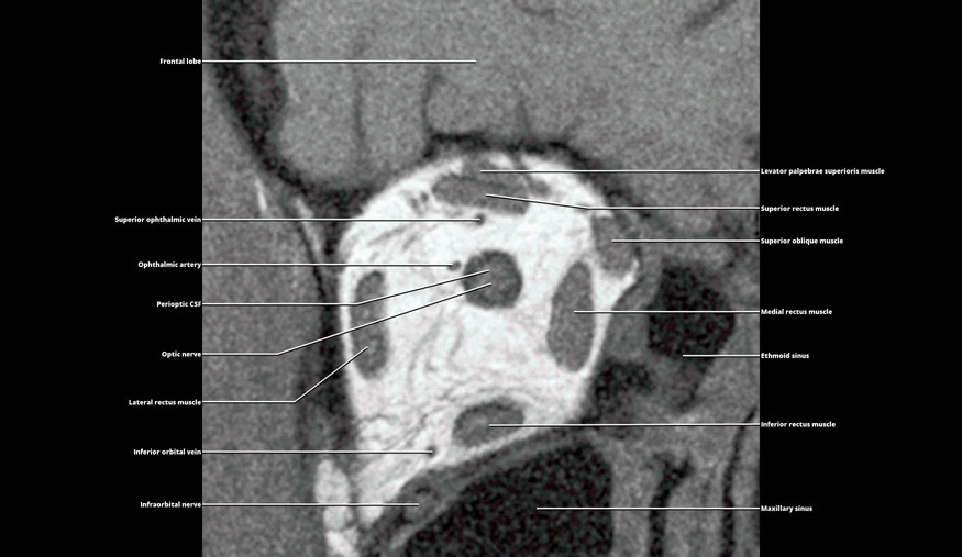
Superior ophthalmic vein
Ophthalmic artery
Perioptic CSF
Optic nerve
Lateral rectus muscle
Inferior orbital vein
Infraorbital nerve
Levator palpebrae superioris muscle
Superior rectus muscle
Superior oblique muscle
Medial rectus muscle
Ethmoid sinus
Inferior rectus muscle
Maxillary sinus
Coronal T1WI MR demonstrates the peripherally located “cone” of extraocular muscles, the central optic nerve sheath complex, and the vascular structures of the orbit. The intrinsic T1 signal of the orbital fat provides excellent contrast for visualizing the intraorbital contents.
Related posts:

Stay updated, free articles. Join our Telegram channel

Full access? Get Clinical Tree


