Middle layer, deep cervical fascia
Tri-color carotid sheath
Deep layer, deep cervical fascia
Pharyngeal mucosal space/surface
Masticator space
Parapharyngeal space
Parotid space
Carotid space
Retropharyngeal space
Perivertebral space
Axial graphic of the normal parapharyngeal space at the level of the nasopharynx demonstrates the complex fascial margins and the fat-only contents. Mass lesions originating in the surrounding pharyngeal mucosal, masticator, parotid, and carotid spaces can extend into the parapharyngeal space. The resulting displacement pattern of the parapharyngeal space may be helpful in defining the space of origin of a mass in the suprahyoid neck.
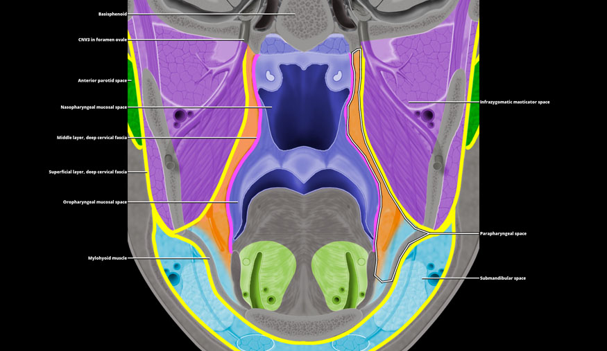
CNV3 in foramen ovale
Anterior parotid space
Nasopharyngeal mucosal space
Middle layer, deep cervical fascia
Superficial layer, deep cervical fascia
Oropharyngeal mucosal space
Mylohyoid muscle
Infrazygomatic masticator space
Parapharyngeal space
Submandibular space
Coronal graphic shows suprahyoid neck spaces as they interact with the skull base superiorly and submandibular space inferiorly. The parapharyngeal space interacts with no critical structures as it abuts the skull base. Inferiorly it empties into the posterior submandibular space along the posterior margin of the mylohyoid muscle. As a consequence of this anatomic arrangement, it is possible for an infection or a malignant tumor that breaks into the parapharyngeal space to present inferiorly as an angle of mandible mass.
Related posts:

Stay updated, free articles. Join our Telegram channel

Full access? Get Clinical Tree


