Spinal Column: Skeletal Pathology
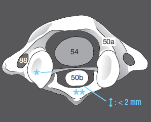
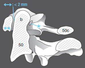
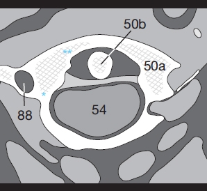
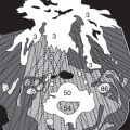
The occipital condyles at the base of the skull articulate with the first vertebra, the atlas (50a), which is the only vertebra to lack a body. The dens (50b) of the axis protrudes upward into the atlas and is held in place by the transverse ligament (*) ( Figs. 152.1 and 152.2 ). This ligament may be torn by a whiplash injury during road traffic accidents.
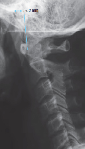
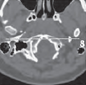
The width of the space (↔) between the anterior arch of the atlas (** in Figs. 152.1 and 152.2 ) and the dens is also measured, as in conventional x-ray images ( Fig. 152.3 ). In adults it should not exceed 2 mm; in children, 4 mm. The vertebral artery passes through the transverse foramen (88).
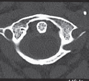
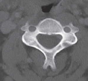
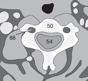
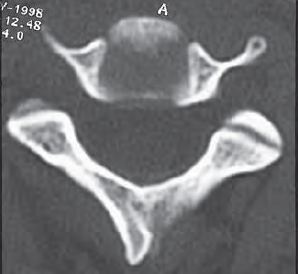
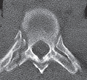
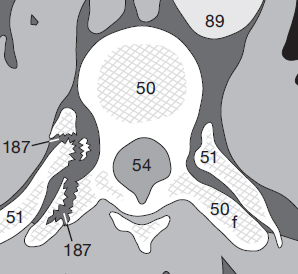
The images below show normal anatomy at the level of the atlas ( Fig. 152.4 ) and the body of the axis ( Fig. 152.5 ). The cartilage of an intervertebral disc (50e in Fig. 152.6 ) will appear more homogeneous and hypodense than the typical pattern of trabeculae.
Cervical Disc Protrusion
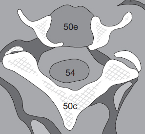
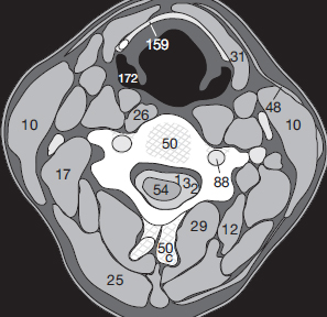
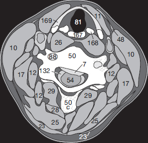
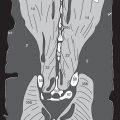
A disc protrusion (prolaps of the nucleus pulposus) is demonstrated optimally in CT sections after myelography (CM in the SAS). The spinal cord is virtually isodense to CSF in unenhanced images, making it difficult to define the contours of the cord. After a myelogram, the CSF (132) will appear hyperdense to the cord (54) as well as to a disc. Normally, the CSF uniformly surrounds the cervical cord ( Fig. 153.1 ). A disc prolapse (7), protruding into the CSF space can be seen because it is hypodense to the opacified CSF ( Fig. 153.2 ). The gap between the cord (54) and vertebral body (50) is filled in. Did you recognize the pyriform fossa (172), the hyoid bone (159), the thyroid cartilage (169), and the cricoid cartilage (167)?
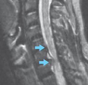
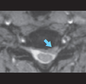
A disc prolapse will be seen even more clearly in an MR image. The sagittal T2-weighted image in Figure 153.3a shows the extent of protrusions at two disk spaces. The disk protrudes into the hyperintense CSF space () in front of the cord. The axial T2-weighted image ( Fig. 153.3b ) shows that the prolapse extends to the left and has caused stenosis of the intervertebral foramen ().
Cervical Spine Fractures
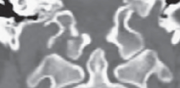
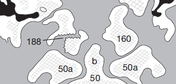
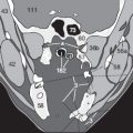
It is especially important to look for fractures of the cervical spine or for torn ligaments after trauma (ref. p. 152) so that damage to the cord is avoided if the patient needs to be moved or transported. Figures 153.4a through c show a coronal MPR in which the right occipital condyle (160) is fractured (188) but the dens (50b) is still in normal position.
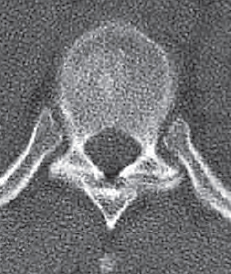
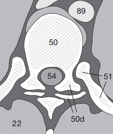
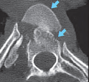
The thoracic vertebrae articulate with each other at their superior and inferior articular facets (50d) and with the ribs (51) at the inferior and superior costal facets and the trans-verse processes (50f). Figure 154.1 shows a normal thoracic image: the contours of the cortical bone are smooth and the trabeculae have a homogeneous pattern.
Related posts:


Stay updated, free articles. Join our Telegram channel

Full access? Get Clinical Tree


