Superficial layer, deep cervical fascia
Middle layer, deep cervical fascia
Retropharyngeal space
Perivertebral space, prevertebral component
Deep layer, deep cervical fascia
Perivertebral space, paraspinal component
Masticator space
Pharyngeal mucosal space/surface
Parapharyngeal space
Parotid space
Carotid space
Axial graphic depicts the spaces of the suprahyoid neck. Surrounding the paired fat-filled parapharyngeal spaces (PPS) are the 4 critical paired spaces of this region, the pharyngeal mucosal (PMS), masticator (MS), parotid (PS), and carotid spaces (CS). Retropharyngeal (RPS) and perivertebral spaces (PVS) are the midline nonpaired spaces. A PMS mass pushes the PPS laterally, an MS mass pushes the PPS posteriorly, a PS mass pushes the PPS medially, and a CS mass pushes the PPS anteriorly. Lateral RPS mass pushes PPS anteriorly without lifting styloid process. The superficial (yellow line), middle (pink line), & deep (turquoise line) layers of deep cervical fascia outline the spaces.
Temporalis muscle
Lateral pterygoid muscle
Styloid process
Internal jugular vein
Internal carotid artery
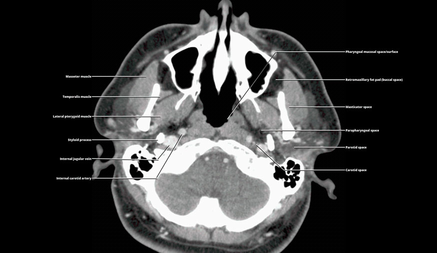
Pharyngeal mucosal space/surface
Retromaxillary fat pad (buccal space)
Masticator space
Parapharyngeal space
Parotid space
Carotid space
Axial CECT at the level of the nasopharyngeal suprahyoid neck shows the 4 key spaces surrounding the PPS: The PMS, MS, PS, and CS. Notice the retropharyngeal fat stripe is not seen in the high nasopharynx between the prevertebral muscles and the pharyngeal mucosal surface.
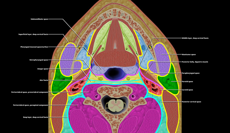
Superficial layer, deep cervical fascia
Pharyngeal mucosal space/surface
Retropharyngeal space
Danger space
Alar fascia
Perivertebral space, prevertebral component
Perivertebral space, paraspinal component
Deep layer, deep cervical fascia
Middle layer, deep cervical fascia
Masticator space
Posterior belly, digastric muscle
Parapharyngeal space
Parotid space
Carotid space
Posterior cervical space
Axial graphic shows the suprahyoid neck spaces at the level of the oropharynx. The superficial (yellow line), middle (pink line), and deep (turquoise line) layers of deep cervical fascia outline the suprahyoid neck spaces. Notice the lateral borders of the RPS & danger spaces are called the alar fascia, which represents a slip of the deep layer of deep cervical fascia. The CS has a tricolored fascial representation for the carotid sheath. This is because all 3 layers of deep cervical fascia contribute to the carotid sheath.
Related posts:

Stay updated, free articles. Join our Telegram channel

Full access? Get Clinical Tree


