Chapter 9 The lumbar muscles and their fasciae
1 Psoas major, which covers the anterolateral aspects of the lumbar spine.
2 Intertransversarii laterales and quadratus lumborum, which connect and cover the transverse processes anteriorly.
3 The lumbar back muscles, which lie behind and cover the posterior elements of the lumbar spine.
Psoas major
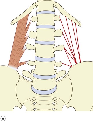
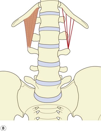
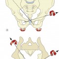
The psoas major has diverse but systematic attachments to the lumbar spine (Fig. 9.1). At each segmental level from T12–L1 to L4–5, it is attached to the medial three-quarters or so of the anterior surface of the transverse process, to the intervertebral disc, and to the margins of the vertebral bodies adjacent to the disc.1 An additional fascicle arises from the L5 vertebral body. Classically, the muscle is also said to arise from a tendinous arch that covers the lateral aspect of the vertebral body.2 Close dissection,1 however, reveals that these arches constitute no more than the medial, deep fascia of the muscle, and that the fascia affords no particular additional origin; the most medial fibres of the muscle skirt the fascia and are anchored directly to the upper margin of the vertebral body. Nonetheless, the fascia forms an arcade deep to the psoas, over the lateral surface of the vertebral body, leaving a space between the arch and the bone that transmits the lumbar arteries and veins (see Ch. 11).
The muscle fibres from the L4–5 intervertebral disc, the L5 body and the L5 transverse process form the deepest and lowest bundle of fibres within the muscle. These fibres are systematically overlapped by fibres from the disc, vertebral margins and transverse process at successively higher levels. As a result, the muscle in cross-section is layered circumferentially, with fibres from higher levels forming the outer surface of the muscle and those from lower levels buried sequentially, deeper within its substance. Within the muscle, bundles from individual lumbar segments have the same length, such that those from L1 become tendinous before those from successively lower levels. This isometric morphology indicates that the muscle is designed exclusively to act on the hip.1
Biomechanical analysis reveals that the psoas has only a feeble action on the lumbar spine with respect to flexion and extension. Its fibres are disposed so as to extend upper lumbar segments and to flex lower lumbar segments. However, the fibres act very close to the axes of rotation of the lumbar vertebrae and so can exert only very small moments, even under maximal contraction.1 This denies the psoas any substantial action on the lumbar spine. Rather, it uses the lumbar spine as a base from which to act on the hip.
However, the psoas potentially exerts massive compression loads on the lower lumbar discs. The proximity of the lines of action of the muscle to the axes of rotation minimises its capacity as a flexor but maximises the axial compression that it exerts. Upon maximum contraction, in an activity such as sit-ups, the two psoas muscles can be expected to exert a compression load on the L5–S1 disc equal to about 100 kg of weight.1
Intertransversarii laterales
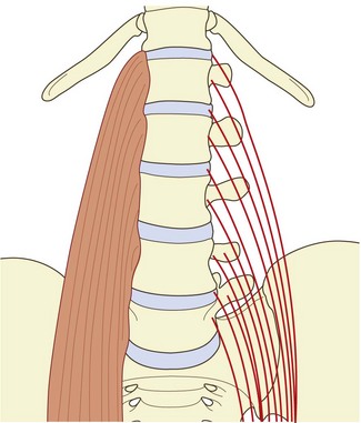
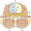
The intertransversarii laterales consist of two parts: the intertransversarii laterales ventrales and the intertransversarii laterales dorsales. The ventral intertransversarii connect the margins of consecutive transverse processes, while the dorsal intertransversarii each connect an accessory process to the transverse process below (Fig. 9.2). Both the ventral and dorsal intertransversarii are innervated by the ventral rami of the lumbar spinal nerves,3 and consequently cannot be classified among the back muscles which are all innervated by the dorsal rami (see Ch. 10). On the basis of their attachments and their nerve supply, the ventral and dorsal intertransversarii are considered to be homologous to the intercostal and levator costae muscles of the thoracic region.3
Quadratus lumborum
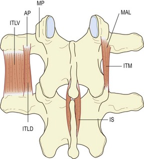
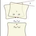
The quadratus lumborum is a wide, more or less rectangular, muscle that covers the lateral two-thirds or so of the anterior surfaces of the L1 to L4 transverse processes and extends laterally a few centimetres beyond the tips of the transverse processes. In detail, the muscle is a complex aggregation of various oblique and longitudinally running fibres that connect the lumbar transverse processes, the ilium and the 12th rib (Fig. 9.3).4
The muscle can be considered as consisting of four types of fascicle arranged in three layers.5 Iliocostal fibres connect the ilium and the 12th rib. Iliolumbar lumbar fibres connect the ilium and the lumbar transverse processes. Lumbocostal fibres connect the lumbar transverse processes and the 12th rib. A fourth type of fascicle connects the ilium and the body of the 12th thoracic vertebra. Occasionally, fascicles may connect the lumbar transverse processes to the body of the 12th thoracic vertebra.
The posterior layer (see Fig. 9.3A) consists of iliolumbar fascicles inferiorly and medially, and iliocostal fascicles laterally.5 The iliolumbar fibres arise from the iliac crest, and most consistently insert into the upper three lumbar transverse processes. Occasionally, some fascicles also insert into the L4 transverse process.
The middle layer (see Fig. 9.3B) typically arises by a common tendon from the anterior surface of the L3 transverse process. Its fascicles radiate to the inferior anterior aspect of the medial half or so of the 12th rib.5 Occasionally these fascicles are joined by ones from the L2, L4 and L5 transverse processes.
The anterior layer (see Fig. 9.3C) consists of more or less parallel fibres stemming from the iliac crest and passing upwards. The more lateral fibres insert into the lower anterior aspect of the 12th rib. More medial fibres insert into a tubercle on the lateral aspect of the body of the 12th thoracic vertebra.5 These latter fascicles may be joined at their insertion by fascicles from the lumbar transverse processes, most often from the L4 and L5 levels, when they occur.
The irregular and inconstant structure of the quadratus lumborum makes it difficult to discern exactly its function. Classically one of the functions of this muscle is said to be to fix the 12th rib during respiration.2 This fits with many, although not most, of its fibres inserting into the 12th rib. The majority of fibres, and the largest, however, anchor the lumbar transverse processes and the 12th thoracic vertebra to the ilium. These attachments indicate that a major action of the muscle would be lateral flexion of the lumbar spine. However, the strength of the muscle is limited by the size of its fascicles and their moment arms. For lateral flexion, the quadratus lumborum can exert a maximum moment of about 35 Nm.5
Since the fascicles of the quadratus lumborum act behind the axes of sagittal rotation of the lumbar vertebrae, they are potentially extensors of the lumbar spine. However, in this role their capacity is limited to about 20 Nm,5 which amounts to less than 10% of the moment exerted by the posterior back muscles.
These limitations in strength leave the actual function of the quadratus lumborum still an enigma.
The lumbar back muscles
1 The short intersegmental muscles – the interspinales and the intertransversarii mediales.
2 The polysegmental muscles that attach to the lumbar vertebrae – the multifidus and the lumbar components of the longissimus and iliocostalis.
3 The long polysegmental muscles, represented by the thoracic components of the longissimus and iliocostalis lumborum, which in general do not attach to the lumbar vertebrae but cross the lumbar region from thoracic levels to find attachments on the ilium and sacrum.
The descriptions of the back muscles offered in this chapter, notably those of the multifidus and erector spinae, differ substantially from those given in standard textbooks. Traditionally, these muscles have been regarded as stemming from a common origin on the sacrum and ilium and passing upwards to assume diverse attachments to the lumbar and thoracic vertebrae and ribs. However, in the face of several studies of these muscles,6–9 it is considered more appropriate to view them in the reverse direction – from above downwards. Not only is this more consistent with the pattern of their nerve supply9,10 but it clarifies the identity of certain muscles and the identity of the erector spinae aponeurosis, and reveals the segmental biomechanical disposition of the muscles.
Interspinales
The lumbar interspinales are short paired muscles that lie on either side of the interspinous ligament and connect the spinous processes of adjacent lumbar vertebrae (see Fig. 9.2). There are four pairs in the lumbar region.
Intertransversarii mediales
The intertransversarii mediales can be considered to be true back muscles for, unlike the intertransversarii laterales, they are innervated by the lumbar dorsal rami.3,10 The intertransversarii mediales arise from an accessory process, the adjoining mamillary process and the mamillo-accessory ligament that connects these two processes.11 They insert into the superior aspect of the mamillary process of the vertebra below (see Fig. 9.2).
A tantalising alternative suggestion is that the intertransversarii (and perhaps also the interspinales) act as large proprioceptive transducers; their value lies not in the force they can exert but in the muscle spindles they contain. Placed close to the lumbar vertebral column, the intertransversarii could monitor the movements of the column and provide feedback that influences the action of the surrounding muscles. Such a role has been suggested for the cervical intertransversarii, which have been found to contain a high density of muscle spindles.12–14 Indeed, all unisegmental muscles of the vertebral column have between two and six times the density of muscles spindles found in the longer polysegmental muscles, and there is growing speculation that this underscores the proprioceptive function of all short, small muscles of the body.15–17
Multifidus
The multifidus is the largest and most medial of the lumbar back muscles. It consists of a repeating series of fascicles which stem from the laminae and spinous processes of the lumbar vertebrae and exhibit a constant pattern of attachments caudally.9
The shortest fascicles of the multifidus are the ‘laminar fibres’, which arise from the caudal end of the dorsal surface of each vertebral lamina and insert into the mamillary process of the vertebra two levels caudad (Fig. 9.4A). The L5 laminar fibres have no mamillary process into which they can insert, and insert instead into an area on the sacrum just above the first dorsal sacral foramen. Because of their attachments, the laminar fibres may be considered homologous to the thoracic rotatores.
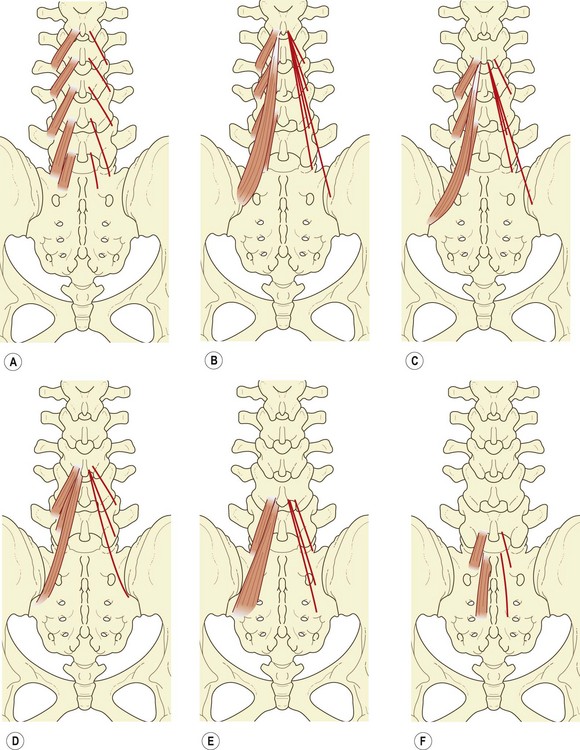
The fascicle from the base of the L1 spinous process inserts into the L4 mamillary process, while those from the common tendon insert into the mamillary processes of L5, S1 and the posterior superior iliac spine (Fig. 9.4B).
The fascicle from the base of the spinous process of L2 inserts into the mamillary process of L5, while those from the common tendon insert into the S1 mamillary process, the posterior superior iliac spine, and an area on the iliac crest just caudoventral to the posterior superior iliac spine (Fig. 9.4C).
The fascicle from the base of the L3 spinous process inserts into the mamillary process of the sacrum, while those fascicles from the common tendon insert into a narrow area extending caudally from the caudal extent of the posterior superior iliac spine to the lateral edge of the third sacral segment (Fig. 9.4D). The L4 fascicles insert onto the sacrum in an area medial to the L3 area of insertion, but lateral to the dorsal sacral foramina (Fig. 9.4E), while those from the L5 vertebra insert onto an area medial to the dorsal sacral foramina (Fig. 9.4F).
It is noteworthy that while many of the fascicles of multifidus attach to mamillary processes, some of the deeper fibres of these fascicles attach to the capsules of the zygapophysial joints next to the mamillary processes (see Ch. 3).18 This attachment allows the multifidus to protect the joint capsule from being caught inside the joint during the movements executed by the multifidus.
The key feature of the morphology of the lumbar multifidus is that its fascicles are arranged segmentally. Each lumbar vertebra is endowed with a group of fascicles that radiate from its spinous process, anchoring it below to mamillary processes, the iliac crest and the sacrum. This disposition suggests that the fibres of multifidus are arranged in such a way that their principal action is focused on individual lumbar spinous processes.9 They are designed to act in concert on a single spinous process. This contention is supported by the pattern of innervation of the muscle. All the fascicles arising from the spinous processes of a given vertebra are innervated by the medial branch of the dorsal ramus that issues from below that vertebra (see Ch. 10).9,10 Thus, the muscles that directly act on a particular vertebral segment are innervated by the nerve of that segment.
In a posterior view, the fascicles of multifidus are seen to have an oblique caudolateral orientation. Their line of action can therefore be resolved into two vectors: a large vertical vector and a considerably smaller horizontal vector (Fig. 9.5A).7
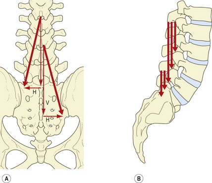
The small horizontal vector suggests that the multifidus could pull the spinous processes sideways, and therefore produce horizontal rotation. However, horizontal rotation of lumbar vertebrae is impeded by the impaction of the contralateral zygapophysial joints. Horizontal rotation occurs after impaction of the joints only if an appropriate shear force is applied to the intervertebral discs (see Ch. 7) but the horizontal vector of multifidus is so small that it is unlikely that the multifidus would be capable of exerting such a shear force on the disc by acting on the spinous process. Indeed, electromyographic studies reveal that the multifidus is inconsistently active in derotation and that, paradoxically, it is active in both ipsilateral and contralateral rotation.19 Rotation, therefore, cannot be inferred to be a primary action of the multifidus. In this context, the multifidus has been said to act only as a ‘stabiliser’ in rotation,18,19 but the aberrant movements, which it is supposed to stabilise, have not been defined (although see below).
The principal action of the multifidus is expressed by its vertical vector, and further insight is gained when this vector is viewed in a lateral projection (Fig. 9.5B). Each fascicle of multifidus, at every level, acts virtually at right angles to its spinous process of origin.7 Thus, using the spinous process as a lever, every fascicle is ideally disposed to produce posterior sagittal rotation of its vertebra. The right-angle orientation, however, precludes any action as a posterior horizontal translator. Therefore, the multifidus can only exert the ‘rocking’ component of extension of the lumbar spine or control this component during flexion.
Having established that the multifidus is primarily a posterior sagittal rotator of the lumbar spine, it is possible to resolve the paradox about its activity during horizontal rotation of the trunk.7 In the first instance, it should be realised that rotation of the lumbar spine is an indirect action. Active rotation of the lumbar spine occurs only if the thorax is first rotated, and is therefore secondary to thoracic rotation. Secondly, it must be realised that a muscle with two vectors of action cannot use these vectors independently. If the muscle contracts, then both vectors are exerted. Thus, the multifidus cannot exert axial rotation without simultaneously exerting a much larger posterior sagittal rotation.
The role of the multifidus in rotation is not to produce rotation but to oppose the flexion effect of the abdominal muscles as they produce rotation. The aberrant motion ‘stabilised’ by the multifidus during rotation is, therefore, the unwanted flexion unavoidably produced by the abdominal muscles.7
Lumbar erector spinae
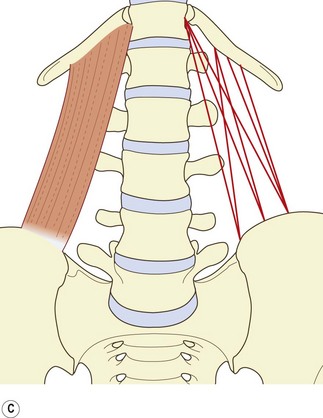
The lumbar erector spinae lies lateral to the multifidus and forms the prominent dorsolateral contour of the back muscles in the lumbar region. It consists of two muscles: the longissimus thoracis and the iliocostalis lumborum. Furthermore, each of these muscles has two components: a lumbar part, consisting of fascicles arising from lumbar vertebrae, and a thoracic part, consisting of fascicles arising from thoracic vertebrae or ribs.6,8 These four parts may be referred to, respectively, as longissimus thoracis pars lumborum, iliocostalis lumborum pars lumborum, longissimus thoracis pars thoracis and iliocostalis lumborum pars thoracis.8
In the lumbar region, the longissimus and iliocostalis are separated from each other by the lumbar intermuscular aponeurosis, an anteroposterior continuation of the erector spinae aponeurosis.6,8 It appears as a flat sheet of collagen fibres, which extend rostrally from the medial aspect of the posterior superior iliac spine for 6–8 cm. It is formed mainly by the caudal tendons of the rostral four fascicles of the lumbar component of longissimus (Fig. 9.6).
Longissimus thoracis pars lumborum
The longissimus thoracis pars lumborum is composed of five fascicles, each arising from the accessory process and the adjacent medial end of the dorsal surface of the transverse process of a lumbar vertebra (see Fig. 9.6).
Each fascicle of the lumbar longissimus has both a dorsoventral and a rostrocaudal orientation.8 Therefore, the action of each fascicle can be resolved into a vertical vector and a horizontal vector, the relative sizes of which differ from L1 to L5 (Fig. 9.7A). Consequently, the relative actions of the longissimus differ at each segmental level. Furthermore, the action of the longissimus, as a whole, will differ according to whether the muscle contracts unilaterally or bilaterally.
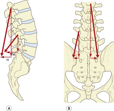
The large vertical vector of each fascicle lies lateral to the axis of lateral flexion and behind the axis of sagittal rotation of each vertebra. Thus, contracting the longissimus unilaterally can laterally flex the vertebral column, but acting bilaterally the various fascicles can act, like the multifidus, to produce posterior sagittal rotation of their vertebra of origin. However, their attachments to the accessory and transverse processes lie close to the axes of sagittal rotation, and therefore their capacity to produce posterior sagittal rotation is less efficient than that of the multifidus, which acts through the long levers of the spinous processes.8
The horizontal vectors of the longissimus are directed backwards. Therefore, when contracting bilaterally the longissimus is capable of drawing the lumbar vertebrae backwards. This action of posterior translation can restore the anterior translation of the lumbar vertebrae that occurs during flexion of the lumbar column (see Ch. 7). The capacity for posterior translation is greatest at lower lumbar levels, where the fascicles of longissimus assume a greater dorsoventral orientation (Fig. 9.7B).
Iliocostalis lumborum pars lumborum
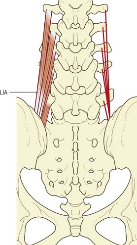
The lumbar component of the iliocostalis lumborum consists of four overlying fascicles arising from the L1 through to the L4 vertebrae. Rostrally, each fascicle attaches to the tip of the transverse process and to an area extending 2–3 cm laterally onto the middle layer of the thoracolumbar fascia (Fig. 9.8).
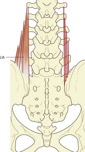
Although an L5 fascicle of iliocostalis lumborum is not described in the literature, it is represented in the iliolumbar ‘ligament’. In neonates and children this ‘ligament’ is said to be completely muscular in structure (see Ch. 4).20 By the third decade of life, the muscle fibres are entirely replaced by collagen, giving rise to the familiar iliolumbar ligament.20 On the basis of sites of attachment and relative orientation, the posterior band of the iliolumbar ligament would appear to be derived from the L5 fascicle of iliocostalis, while the anterior band of the ligament is a derivative of the quadratus lumborum.
The disposition of the lumbar fascicles of iliocostalis is similar to that of the lumbar longissimus, except that the fascicles are situated more laterally. Like that of the lumbar longissimus, their action can be resolved into horizontal and vertical vectors (Fig. 9.9A).
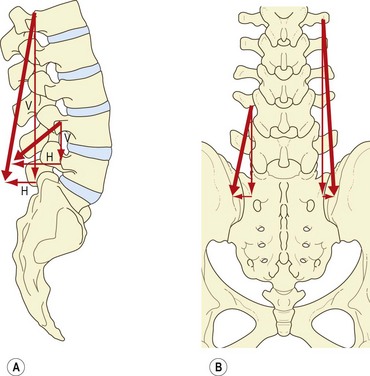
The vertical vector is still predominant, and therefore the lumbar fascicles of iliocostalis contracting bilaterally can act as posterior sagittal rotators (Fig. 9.9B), but because of the horizontal vector a posterior translation will be exerted simultaneously, principally at lower lumbar levels where the fascicles of iliocostalis have a greater forward orientation. Contracting unilaterally, the lumbar fascicles of iliocostalis can act as lateral flexors of the lumbar vertebrae, for which action the transverse processes provide very substantial levers.
Stay updated, free articles. Join our Telegram channel

Full access? Get Clinical Tree


