Key Points
- •
The four main types of ultrasound transducers—linear, curvilinear, phased-array, and intracavitary—differ by crystal arrangement, size, and footprints, which determine their suitability in different imaging applications.
- •
Higher-frequency transducers produce high-resolution images of superficial structures, whereas low-frequency transducers produce low-resolution images of deep structures.
- •
Resolution of ultrasound images is divided into four different types: axial, lateral, elevational, and temporal.
Background
Ultrasonography utilizes the piezoelectric effect , or ability of certain crystals to generate vibrations with the application of electricity. The vibrating crystals, also known as piezoelectric elements, generate ultrasonic waves that are transmitted to an apposed object. Reflected waves return to the transducer and generate mechanical distortion of the crystals, which is converted to an electric current via the same piezoelectric effect. The electric current is interpreted by the ultrasound machine’s computer processor and rendered into an image. Transducers are a fundamental component of ultrasound imaging. Point-of-care ultrasound users should have a basic understanding of transducer characteristics, including types and construction, and an understanding of determinants of image resolution.
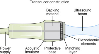
Transducer Construction
Ultrasound transducers are designed for optimal transmission and reception of sound waves ( Figure 3.1 ). An electrical shield lines the transducer case to prevent external electrical interference from distorting sound wave transmission. A thin acoustic insulator dampens vibrations from the case to piezoelectric elements and prevents transmission of spurious electric current to the machine’s computer processor. At the tip of the transducer, a thin matching layer improves efficiency of sound wave transmission from piezoelectric elements to skin and deeper structures. Backing material is an essential component of transducers. Backing material is fixed behind the layer of piezoelectric elements to dampen ongoing vibrations of elements. Sound energy is absorbed by backing material when elements are generating and receiving sound waves. Transducers are sensitive instruments, and the internal components of the transducer head, including the piezoelectric elements, can break easily with minor impact. Providers must be trained to safeguard transducers at all times.
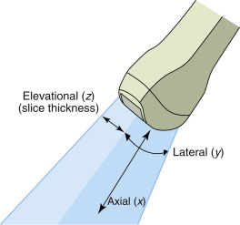
Resolution
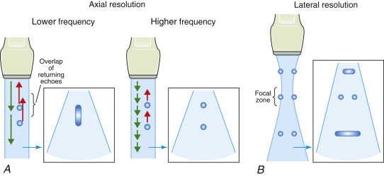
Resolution of ultrasound images depends on three complementary properties of the transducer: axial, lateral, and elevational resolution ( Figure 3.2 ). Axial resolution is the ability to differentiate distinct objects on the same path as the ultrasound beam. Lateral resolution is the ability to differentiate objects that are perpendicular to the ultrasound beam. Axial resolution is determined by sound wave frequency, with higher frequencies giving better axial resolution ( Figure 3.3 A ). Lateral resolution is determined by width of the ultrasound beam, which is influenced by diameter and frequency of piezoelectric crystals. Small-diameter crystals that produce high-frequency pulses generate narrow ultrasound beams, and thereby increase lateral resolution. More important, the narrowest portion of the ultrasound beam, or focal zone, has the greatest lateral resolution. Depth of the focal zone can be adjusted to the level of the target structure to maximize lateral resolution ( Figure 3.3 B). Ultrasound computer processors adjust sound wave frequency to maximize axial and lateral resolution when depth settings are changed.