Multiple regenerating nodules (NECT or MR), same as other cirrhotic nodules
• CT and MR for evaluation of cirrhosis and HCC
Not for diagnosis of Wilson disease
There are no specific imaging features for hepatic injury due to Wilson disease
TOP DIFFERENTIAL DIAGNOSES
• Steatosis (fatty liver)
• Hepatitis
PATHOLOGY
• Steatosis → fibrosis and ultimately cirrhosis
CLINICAL ISSUES
• Presentation: Chronic hepatitis, cirrhosis, acute liver failure in adolescent or young adult
• Acute fulminant presentation of Wilson disease most often in females (M:F = 1:2)
• Prevalence: 1 in 30,000 (not rare)
• Diagnosis: Liver biopsy and copper quantitation
Presence of Kayser-Fleisher rings and low level of ceruloplasmin sufficient to diagnose Wilson disease
• Therapy: Copper chelation or transplantation
Those with acute hepatic failure have worse prognosis
Initial and maintenance therapy with copper chelation
Liver transplantation cures the disease and prevents recurrence
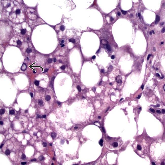
(Left) Hematoxylin & eosin stained section shows features of steatohepatitis in Wilson disease (WD) with steatosis and glycogenated nuclei . (Courtesy J. Misdraji, MD.)
(Right) Aldehyde fuchsin stain shows darkly staining granules of copper-associated protein (metallothionein) in periportal hepatocytes . (Courtesy J. Misdraji, MD.)
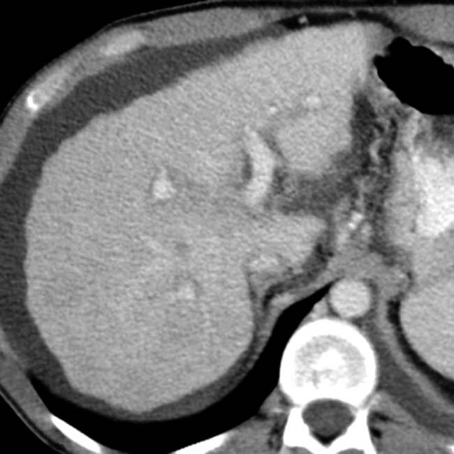
(Left) Axial NECT shows a nodular shrunken liver, typical of cirrhosis. Within the liver are innumerable hyperdense nodules that are typical of cirrhotic regenerating nodules, which are not necessarily indicative of Wilson disease.
(Right) Axial venous phase CECT in the same patient shows that the regenerating nodules become nearly isodense with the liver, as usual. Signs of portal hypertension are evident, including ascites and varices. Cirrhosis was due to Wilson disease in this patient.
TERMINOLOGY
Abbreviations
• Wilson disease (WD)
Synonyms
• Hepatolenticular degeneration
Definitions
• Autosomal recessive disorder in which copper (Cu) accumulates pathologically
Primarily within liver and subsequently in neurologic system and other tissues
IMAGING
General Features
• Best diagnostic clue
Early onset of cirrhosis, but no specific imaging features
• Location
Early: Diffuse distribution of copper in liver cytoplasm
Later: Within lysosomes, then throughout liver nodules
• Key concepts
There are no specific imaging features for hepatic injury due to Wilson disease
CT Findings
• Spectrum of hepatic injury nonspecific; changes of fatty infiltration or cirrhosis frequently indistinguishable from other etiologies
• Copper has high atomic number and can cause elevation of liver density on CT
Unusual finding because coexisting fatty infiltration diminishes hepatic parenchymal attenuation
• Multiple hyperdense regenerating nodules (on NECT)


 . (Courtesy J. Misdraji, MD.)
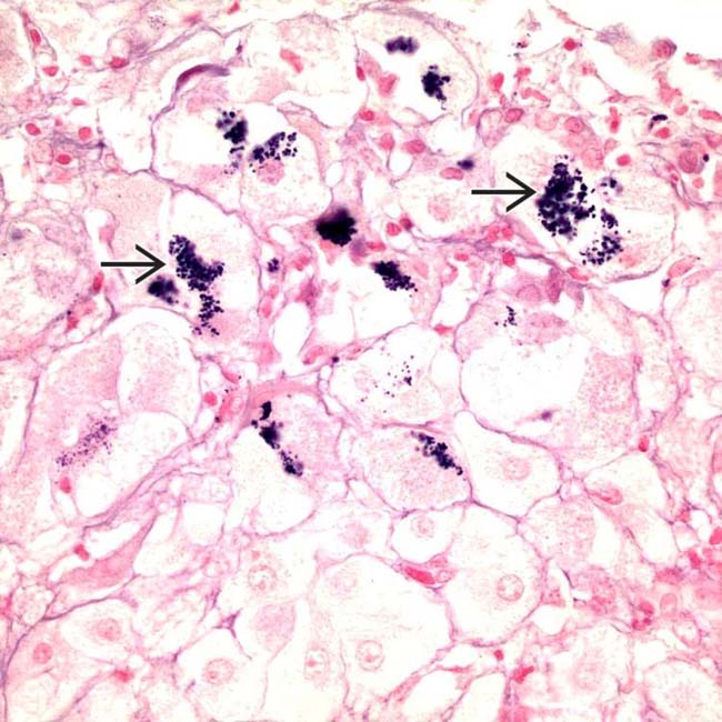
 . (Courtesy J. Misdraji, MD.)
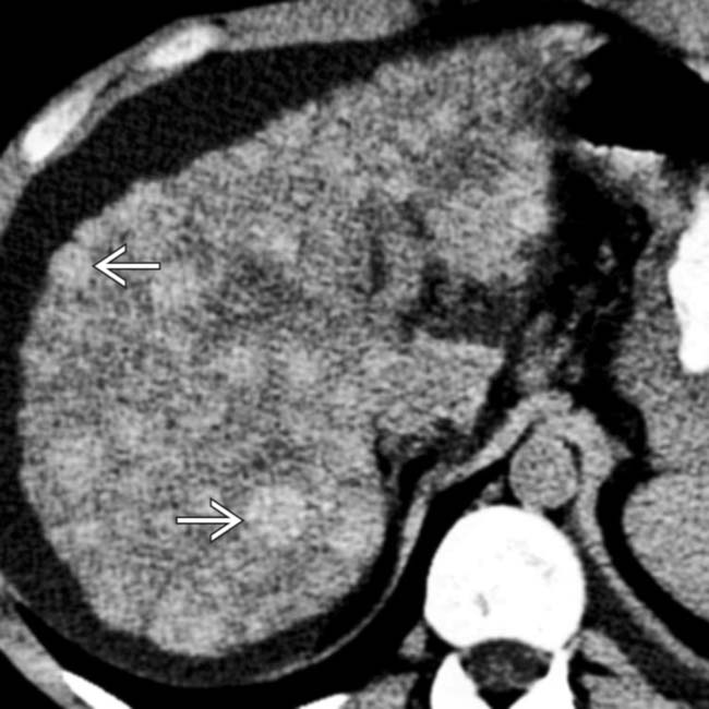
 that are typical of cirrhotic regenerating nodules, which are not necessarily indicative of Wilson disease.