1.2 Image Interpretation
Before any remarkable image details are interpreted, they must be described. This does not require a lengthy narrative but may be done concisely in a “key-word” format. The description of findings should nevertheless be exacting and, above all, should avoid potentially misleading language. For example, a lucent area on a radiograph or CT scan should be described as such and should not be called a “cystic” or “cystlike” osteolysis, as this could bias further conclusions that may be drawn in the light of other findings. Another example is “bone marrow edema,” a term that is frequently overused in MRI reporting (see below).
Note
Image analysis requires a detailed knowledge of the normal radiologic anatomy of the musculoskeletal system and its principal variants.
1.2.1 Pathoanatomic Background
When a finding is classified as definitely abnormal, the first essential question should address its possible pathoanatomic background, because each of the known basic entities (see below) is associated with a more or less specific pathoanatomic change. For example, a tumor or inflammatory process is associated with destruction, necrosis with fragmentation, and a reparative process with new bone formation. An effort should also be made to determine whether a finding may be only an epiphenomenon (e.g., hemorrhage, callus) that accompanies an underlying process. In assigning findings to a pathoanatomic substrate, it is always important to consider the capabilities for tissue discrimination (fat, fluid, etc.) that are available with the modality being used. Unfortunately, this capability is often forgotten in the case of CT (see Case 58).
In the process of image interpretation, it is dangerous to use terms that describe either a disease entity in the strict sense or a sign with a variegated background. This principle will be explained more fully based on the two examples mentioned above:
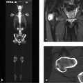
Example: Rounded Lucency
In many cases a rounded lucency seen on a radiograph or CT scan is described as a “cyst” or a “cystic or cystlike lucency,” even though it is more often caused by a solid destructive lesion. This wording may steer case deliberations still further in the wrong direction, implying that the finding may be innocent. The term “cyst” is reserved exclusively for a fluid-filled cavity such as a juvenile bone cyst, a subchondral synovial cyst, or an aneurysmal bone cyst. The fluid content should first be established by CT or MRI, except with a classic juvenile bone cyst, which is identified by its typical location, “fallen fragment sign,” and ultrashort history.
Example: Ill-defined Hyperintensity
An area of increased signal intensity with ill-defined margins seen in water-sensitive MRI sequences is often described simply as “edema.” This is frequently offered as a diagnosis when no other suggestive findings are seen. Edema in bone marrow or in structurally altered cortical bone is an extravascular interstitial fluid collection that may accompany, for instance, a traumatic, inflammatory, degenerative or even a neoplastic process. “Edema” is a term that runs the gamut from benign to malignant disorders. Simply put, the resulting MRI signal is based partly on an increased proton density in the imaged region, regardless of the cause, which is usually but not always edema (see below).
Based on the MR image alone, it cannot be stated with certainty whether “bone marrow edema” actually represents pure edema, meaning an extravascular interstitial fluid collection. Taking degenerative changes in the knee joint as an example, “bone marrow edema” may result from fat necrosis, fibrosis, or trabecular bone changes such as a bone bruise, as Zanetti et al12 have shown. A pathologic process need only have enough protons to cause increased signal intensity in water-sensitive sequences. This applies to traumatic, inflammatory, degenerative, and neoplastic processes, the latter also showing an increased intracellular water content (see Case 145, Case 146, and Case 149). Even contrast administration cannot positively distinguish, for example, between tumor tissue and edema. It is clearer and more accurate, therefore, to report an edema equivalent13 , 14 or an “edema-like hyperintensity,” or simply to report the presence of a proton-rich area.
1.2.2 Lesion Localization
The second question raised by an abnormal finding is its location, which is considered in terms of functional anatomy. Is the finding located in a stress-exposed area (e.g., entheses), a critical area for perfusion deficits (e.g., epiphyses, toes or fingers), or a region where hematopoiesis occurs (e.g., a vertebral body)? These are key questions that aid in the classification of findings.
1.2.3 Epicenter of a Lesion
The third question relates to the epicenter of a lesion or other finding. Most neoplastic processes exhibit a concentric pattern of tumor growth. Thus, for example, if the center of a lesion is located in the cortex, it is reasonable to assume that the process originated in the cortex or periosteum (cortical osteoid osteoma, periosteal osteosarcoma, stress fracture, etc.). If there is an enthesis in the affected area, it should also be considered that stress at the insertion site may have led to cortical destruction or new bone formation (see Case 103 and Case 104).
Related posts:

Stay updated, free articles. Join our Telegram channel

Full access? Get Clinical Tree


