4.5 Soft-Tissue Mineralization
4.5.1 Case 66 (Fig. 4.29)

Case description
Referring physician: nephrologist.
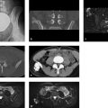
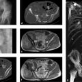
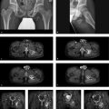
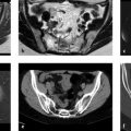
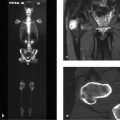
Prior history and clinical question: This patient, already on long-term dialysis, was 24 years old when the images shown here (Fig. 4.29) were obtained. His hyperphosphatemia due to chronic renal failure had not been treated for reasons not detailed here. The patient recently developed swellings about the shoulder, hip, and several metacarpophalangeal joints. The nephrologist wanted to know the cause of the joint swellings.
Radiologic Findings
Radiographs show extensive mineralized masses of calcific density (“calcifications”) about the hip joints, especially on the right side (Fig. 4.29 a), on the ulnar side of the right fourth metacarpophalangeal joint (Fig. 4.29 b), and above the olecranon (Fig. 4.29 c). There were also grotesque calcifications about the shoulder joints (not pictured here). Coarsening of the trabeculae is noted in the bones of the hand, and the bony margins of the terminal tufts are ill defined (Fig. 4.29 b).
Related posts:

Stay updated, free articles. Join our Telegram channel

Full access? Get Clinical Tree


