Abstract
Hippocampal encephalitis (HE), or more generally limbic encephalitis, can be secondary to various etiologies. Through this case, we report a very rare toxic cause: acute hippocampal encephalopathy secondary to cannabis use in a heavy cannabis user (>20 joints/day). A 39-year-old male presented with a feverless disturbance of consciousness. The MRI revealed signal abnormalities in the hippocampal regions. Further investigations ruled out other causes of encephalitis. No renal function impairment or rhabdomyolysis was found. The objective of our study is to describe the radiological features of this severe neurological complication of cannabis
Case report
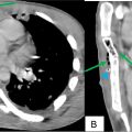
We report the case of a 39-year-old man from a low socio-economic background, living in an urban environment. He had no significant medical or surgical history, with a history of smoking, though the extent was not quantified. He presented with a feverless disturbance of consciousness. His family reported excessive cannabis consumption, with a daily intake of over 20 joints. Clinical evaluation revealed a patient with a Glasgow Coma Scale (GCS) of 10/15, without focal neurological signs. No cardio-respiratory abnormalities were noted. Blood glucose testing showed no glycemic abnormalities. The patient was afebrile. Biological tests revealed a mild inflammatory syndrome with white blood cells at 17,000/mm³. Cerebrospinal fluid analysis was normal, with no signs of meningitis or antibodies linked to autoimmune encephalitis. Toxicological screening confirmed the presence of cannabis in the bloodstream, with no other toxic substances found. An urgent brain MRI revealed bilateral and symmetric signal abnormalities in the hippocampi on T2 and FLAIR sequences, with restricted diffusion ( Figs. 1–3 ). No pathological contrast enhancement was observed ( Fig. 4 ). Hippocampal volume was slightly reduced. Based on all these findings, a diagnosis of acute hippocampal encephalopathy secondary to cannabis was established. After a stay in medical intensive care and stabilization, the patient’s condition improved, with noticeable improvement in his consciousness disturbance a few days after diagnosis.


Related posts:
 Breast cancer with medullary features shows a fast and plateau enhancement pattern on magnetic resonance images: A case report
 A rare and life-threatening case of spontaneous hemopneumothorax presenting with severe hemodynamic instability
 Postextubation negative-pressure pulmonary edema after an appendectomy
 Primary sternal osteomyelitis in an immunocompetent adolescent
 Avascular necrosis of lateral femoral condyle in a middle-aged woman from central India
 Facial nerve palsy from temporal bone metastasis in papillary carcinoma of thyroid: A case report
Stay updated, free articles. Join our Telegram channel

Full access? Get Clinical Tree


