Abdominal Mesothelioma
Michael P. Federle, MD, FACR
Key Facts
Terminology
Rare primary malignant neoplasm arising from peritoneum
Imaging
70% of malignant mesotheliomas arise in pleura
20-30% of malignant mesotheliomas arise in peritoneum
May also arise in pericardium, tunica vaginalis
Calcified pleural plaques in 50% of patients with peritoneal mesotheliomas
Omental and peritoneal-based masses
Stellate, thickened (“pleated”) mesentery secondary to encasement and straightening of mesenteric vessels
Spreads along serosal surfaces and directly invades adjacent viscera, especially colon and liver
Variable amount of ascites; massive ascites uncommon
Top Differential Diagnoses
Peritoneal metastases and lymphoma
Cannot be distinguished from mesothelioma by imaging
Is much more common than mesothelioma
Pseudomyxoma peritonei
Peritoneal inclusion cyst
Sometimes (incorrectly) called “benign cystic (or multicystic) mesothelioma”
Has nothing in common with malignant mesothelioma
Tuberculosis, abdominal manifestations
Pathology
Associated with asbestos exposure
20-40 year latency between exposure and diagnosis
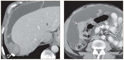 (Left) Axial CT in an elderly man with abdominal distention shows a calcified pleural asbestos plaque  . The parietal peritoneum under the diaphragm is diffusely thickened . The parietal peritoneum under the diaphragm is diffusely thickened  . (Right) Axial CECT in the same patient shows an omental mass . (Right) Axial CECT in the same patient shows an omental mass  , loculated ascites, and a “stiff” mesentery (better shown on other sections). The abdominal findings are indistinguishable from peritoneal carcinomatosis, but the asbestos plaque is an important clue to the diagnosis of mesothelioma. , loculated ascites, and a “stiff” mesentery (better shown on other sections). The abdominal findings are indistinguishable from peritoneal carcinomatosis, but the asbestos plaque is an important clue to the diagnosis of mesothelioma. |
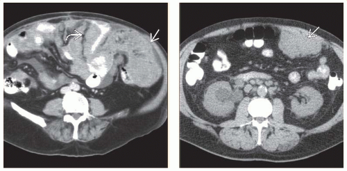 (Left) Axial CECT in an elderly man with abdominal pain shows marked omental thickening  and encasement of bowel loops and encasement of bowel loops  . Open biopsy confirmed malignant mesothelioma. (Right) Axial NECT in a patient with renal insufficiency shows a lobulated mass . Open biopsy confirmed malignant mesothelioma. (Right) Axial NECT in a patient with renal insufficiency shows a lobulated mass  along the peritoneal surface. Surgical biopsy confirmed malignant mesothelioma. This isolated mass is an unusual manifestation of the disease, which is usually widespread at the time of diagnosis. along the peritoneal surface. Surgical biopsy confirmed malignant mesothelioma. This isolated mass is an unusual manifestation of the disease, which is usually widespread at the time of diagnosis. |
TERMINOLOGY
Abbreviations
Malignant mesothelioma (MM), peritoneal mesothelioma (PM)
Definitions
Primary malignant neoplasm arising from peritoneum
“Benign cystic mesothelioma” is misnomer
More accurately referred to as “peritoneal inclusion cyst”
Has nothing in common with malignant mesothelioma
Other than having mesothelial cells in its lining
IMAGING
General Features
Best diagnostic clue
Peritoneal masses or omental cake associated with calcified pleural plaques
Location
70% of malignant mesotheliomas arise in pleura
20-30% of malignant mesotheliomas arise in peritoneum
Few involve both pleura and peritoneum
May also arise in pericardium, tunica vaginalis
Size
Usually involves peritoneal surfaces diffusely or multifocally
Focal masses from few mm to many cm
Morphology
2 primary forms
Desmoplastic form: Diffuse disease thickening peritoneal surfaces and enveloping viscera
Focal form: Large tumor mass with scattered satellite peritoneal nodules
Radiographic Findings
Related posts:

Stay updated, free articles. Join our Telegram channel

Full access? Get Clinical Tree


