Abstract
Objective
Screening ultrasound for hepatocellular carcinoma (HCC) identifies lesions which require further characterization by a contrast-enhanced exam to non-invasively diagnose HCC. While ultrasound is recommended in screening, some HCC can be occult on grayscale imaging. The purpose of this study was to determine if the addition of ultrasound contrast (sulfahexafluoride) to screening ultrasound for HCC can identify more HCC lesions than grayscale sonographic imaging alone.
Methods
All HCC screening ultrasounds that also had contrast were evaluated in this retrospective study. Patients with a focal lesion seen only after administration of contrast (OAC) were noted, as well as any follow-up imaging or pathology results. Additional variables collected included patient demographics, cirrhosis type, and laboratory values.
Results
230 unique patients were included, of which 160 had imaging or pathology follow-up. 18 of these patients had an OAC lesion, of which 17 had follow-up. Among these OACs, there was one LIRADS M lesion (1/18, 5.6 %) and one bland portal vein thrombus identified, which were both confirmed on follow-up imaging. All LIRADS 4 OAC lesions were downgraded. No additional HCC were identified on follow-up imaging or pathology of these patients.
Conclusion
Addition of contrast to screening ultrasound did identify additional lesions, portal vein thrombus, and high grade malignancy. However, as the incidence of OAC lesions was low (7.8 %, 18/230) and most of the lesions were not malignant, addition of post contrast sweeps through the liver is of low value in the low to medium at-risk cirrhotic population in identifying occult HCC.
Graphical abstract

Highlights
- •
Ultrasound screening for hepatocellular carcinoma (HCC) identifies lesions that require further characterization by a contrast-enhanced exam – CT, MRI, or contrast ultrasound (CEUS) to non-invasively diagnose HCC. Screening by ultrasound is recommended but some institutions utilize multi-phase CT or MRI to screen as ultrasound could miss isoechoic lesions.
- •
This study looked to determine if the addition of contrast to screening ultrasound for HCC can identify more lesions than grayscale ultrasound alone and if these OAC lesions (only-after-contrast) increase sensitivity of screening by ultrasound by diagnosing more malignancies.
- •
The rate of identification of OAC during the study was low, and most intermediate LIRADS lesions were downgraded (LIRADS 3 and 4). It was accurate in identifying one ultrasound-occult malignancy and a portal vein thrombus, which were otherwise isoechoic.
- •
Clinical impact.
- •
The addition of contrast to all screening US is low value, but may still be considered in very high risk patients in the cirrhotic at-risk population if there are contraindications to CT or MRI.
1
Introduction
Hepatocellular carcinoma (HCC) is the fourth leading cause of cancer-related mortality worldwide [ , ]. In patients with HCC, early diagnosis is paramount – five-year survival rates are greater than 50 % when detected early, while the detection after tumor symptoms develop is associated with a survival rate of only 10 % . HCC detected at an earlier stage is amenable to liver transplantation or locoregional treatments such as trans-arterial chemoembolization or chemoradiation, radiation therapy, or ablation, which are associated with the best survival outcomes .
The gold standard non-invasive imaging screening options for HCC are multiphase computed tomography (CT) or magnetic resonance imaging (MRI) , however these may not be appropriate or approved by insurance for all at-risk patients. Limitations include allergic reactions to iodinated contrast used in CT, which are less common in CEUS, hesitancy of using iodinated contrast in patients with significantly impaired renal function , and non-diagnostic imaging in the presence of patient motion or breathing. In the screening population, access to accurate alternate imaging is paramount, of which ultrasound is readily available. Ultrasound is recommended as an initial screening modality in the American Association for the Study of Liver Disease (AASLD) guidelines, which recommend ultrasound every six months for high risk patients, with CT or MRI performed if ultrasound is inadequate [ , ].
During ultrasound screening, patients with any indeterminate or suspicious focal liver lesions require a return appointment for contrast-enhanced imaging to characterize the lesion and determine if it meets imaging diagnosis of HCC. Contrast enhanced ultrasound (CEUS) offers accurate [ , , ] and non-invasive diagnosis of HCC for which Liver Imaging-Reporting and Data System (LIRADS) criteria can be applied , based on its arterial and washout characteristics.
Advantages of CEUS include its ability to allow for the real-time monitoring of enhancement and washout. Late phase arterial enhancement, which is important in non-invasive diagnosis of HCC, starts and ends rapidly, and if not optimally timed, may be missed on CT or MRI where imaging timepoints are fixed . CEUS contrast agents remain intravascular and so allows for re-administration if initial characterization is inconclusive or characterization of additional lesions is necessary. However, both non-contrast and CEUS ultrasound can be limited by body habitus and degree of hepatic steatosis.
Screening ultrasound is generally performed without contrast. Ultrasound-occult HCC is a concern [ , ], which can be due to incomplete visualization of the liver parenchyma, background heterogeneity of the liver in cirrhotic patients, isoechoic lesions, or lack of a dedicated imaging protocol. Adding contrast when unenhanced ultrasound does not demonstrate any suspicious lesion has the potential to identify such occult lesions over background liver [ , ]. A screening protocol for HCC utilizing grayscale and CEUS has been described , and has been evaluated internationally using longer-acting Sonozoid (perfluorobutane), a contrast medium not currently Food and Drugs Administration (FDA)-approved in the United States [ , , ]. Park et.al. found that the addition of Sonozoid reduced the false positive rate but did not improve detection rate of early-stage HCC . A multi-institutional trial by Kudo et.al. however, found the addition of CEUS to be useful in identifying early HCC using a reinjection technique . It is not known if Lumason® (sulfahexafluoride), a shorter acting agent could be effective in a more heterogeneous US population.
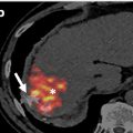
We postulate that lesions identified only after contrast administration (OAC) may be visible during post contrast sweeps through the liver – by their rapid enhancement, rapid washout, and/or on the delayed washout phase. This retrospective study looks at all OAC lesions found during screening ultrasound performed after sulfahexafluoride administration, to determine if the addition of ultrasound contrast in the setting of HCC screening by ultrasound has added value in identifying occult HCC.
2
Methods and materials
With institutional review board (IRB) approval and informed consent waiver, a retrospective study via chart review was conducted at our 546 bed semi-rural institution. All ultrasounds performed for HCC screening from January 2019 through December 2020 were identified. Subjects were included if they were (1) over the age of 18 and (2) they had a liver ultrasound looking for HCC and had contrast. Patients with a confirmatory follow-up test (multiphase CT, MRI, histologic, follow-up screening US) were included. Histologic confirmation included biopsy results, liver explantation, and autopsy results. Exclusion criteria included (1) CEUS ordered for a non-HCC liver lesion evaluation, (2) those who had CEUS for any other reason other than “screening for HCC.”
2.1
Imaging protocol
The screening protocol utilized consisted of grayscale images to evaluate for focal liver lesion or portal vein thrombus, as well as cine sweeps. Post contrast sweeps were performed from 0 to 1 min and from 3 to 4 min to look for enhancement and washout (early and delayed). Contrast microbubbles were then burst, and a second dose through the remaining liver lobe was performed similarly . Images were acquired on Siemens or GE ultrasound units. Amount of sulfahexafluoride contrast was dependent on the machine used, with less required with newer software (1 mL + 5 mL saline flush for newer machines, and 2.4 mL + 5 mL saline flush for older machines).
2.2
Data collection
Variables collected include patient demographics (age, gender, height and weight, type of cirrhosis), information regarding the CEUS scan (enhancement and washout characteristics, LIRADS scores when applicable, portal vein patency, and OAC lesions), laboratory values (alpha-fetoprotein levels, MELD), and follow-up imaging or pathology results/concordance. Two fellowship-trained abdominal radiologists retrospectively reviewed the report and images for OAC (>10 years, and 8 years of experience respectively). These readers checked LIRADS designations and if not present, assigned them based on imaging criteria.
3
Results
3.1
Demographics
A total of 230 HCC screening ultrasounds using contrast were performed during the time period. Of these, 62.6 % (144/230) were men. Mean age at time of CEUS was 59.6 years (range of 20–85). Additional demographics are detailed in Table 1 .
| Patients | |
|---|---|
| Characteristic | ( N = 230) |
| Age (mean ± SD) | 60 ± 13 |
| Gender (M/F) | 144/86 |
| Ethnicity | |
| American Indian/Alaska native | 0 |
| Asian | 16 |
| Native Hawaiian/other Pacific Islander | 0 |
| Black or African American | 13 |
| White/Caucasian | 180 |
| More than one race | 3 |
| Unknown | 18 |
| Height | 170 ± 10 |
| Weight | 91 ± 25 |
| BMI | 31 ± 7 |
| Cirrhosis Etiology | |
| Hepatitis B | 19 |
| Hepatitis C | 45 |
| NASH vs. NAFLD | 84 |
| Alcohol | 52 |
| Cardiogenic | 12 |
| Cryptogenic | 6 |
| Other | 41 |
| AFP baseline | 4.4 ± 7.2 |
| MELDNa baseline | 11 ± 6 |
Related posts:
 Feasibility of deep learning-reconstructed thin-slice single-breath-hold HASTE for detecting pancreatic lesions: A comparison with two conventional T2-weighted imaging sequences
 Non-visualization of axillary pathological lymph nodes in breast cancer patients on SPECT/CT and during operation
 Development of a deep learning model for the automated detection of green pixels indicative of gout on dual energy CT scan
 FDG uptake of pulmonary lesions in synchronous primary lung cancers and lung metastases
 Combination of intrahepatic TARE and extrahepatic TACE to treat HCC patients with extrahepatic artery supply: A case series
 Impact of inspiration level on lung nodule volumetry
Stay updated, free articles. Join our Telegram channel

Full access? Get Clinical Tree


