Adenocarcinoma Mimicking Pneumonia
Katherine R. Birchard
CLINICAL HISTORY
55-year-old female with fatigue.
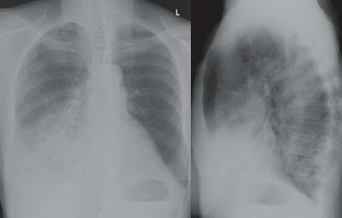 FIGURE 2A |
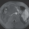
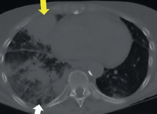 FIGURE 2B |
FINDINGS
Figure 2A: Posteroanterior and lateral chest films show heterogeneous opacities in the right middle and right lower lobes, silhouetting the right heart border and right hemidiaphragm. Normal right hilar structures are obscured. Lateral film shows opacities in the middle lobe are denser than in the right lower lobe. Thickening of right minor and major fissures also evident. Figure 2B: Axial CT image of the lungs shows dense focal opacity in right middle lobe (yellow arrow) and less dense heterogeneous opacities in right lower lobe (white arrow). There is also thickening and nodularity of the major fissure in between the two lobes.
Related posts:


Stay updated, free articles. Join our Telegram channel

Full access? Get Clinical Tree


