KEY FACTS
Terminology
- •
Adnexal torsion is more accurate term than ovarian torsion, as torsion usually also includes fallopian tube
Imaging
- •
Ovary > 4 cm long or > 20 cm³ in volume
- •
Enlarged, heterogeneously echogenic ovarian stroma
- •
Multiple small, fluid-filled follicles displaced peripherally due to edematous stroma &/or mass
- ○
May see follicular ring sign: Thin echogenic rim around follicles in early torsion
- ○
- •
Whirlpool sign: Coiled, twisted pedicle
- •
Flow pattern depends on degree of vascular obstruction and chronicity of torsion
- •
Venous flow affected 1st
- •
Due to dual arterial blood supply to ovary, arterial flow may be preserved
- •
CT
- ○
Twisted pedicle most specific sign but seen in < 1/3 of cases (use multiplanar reformations)
- ○
Heterogeneous, minimal, or absent enhancement indicates evolution from ischemia to infarction
- ○
Top Differential Diagnoses
- •
Hemorrhagic corpus luteum
- •
Pelvic inflammatory disease
- •
Ectopic pregnancy
Pathology
- •
In adults, 50-90% have associated ovarian mass that serves as lead point
- ○
Large physiologic follicular cyst or corpus luteum cyst most common, followed by dermoid
- ○
- •
Presence of venous flow suggests viable ovary
Scanning Tips
- •
Compare with asymptomatic contralateral side
- •
Presence of normal blood flow does not exclude torsion
- •
Always look for underlying mass, which can act as lead point for torsion
- •
Ovaries are highly mobile and when torsed, may be located in unusual locations
- •
When ovaries are deep in location, it may be difficult to detect color Doppler flow even in normal ovaries
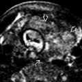
 containing a hemorrhagic cyst with multiple peripheral follicles
  . One of the follicles demonstrates the follicular ring sign
  .
.

 with no color Doppler signal. Alternating red and blue color signal
  represents a twisted vascular pedicle, which is best seen during real time.
 .
.
 in the left adnexal mass with associated hemoperitoneum
  (note the uterus
  ).
).
 and large, bluish hemorrhagic cyst
  .
.
 . Blood flow is seen
  , and despite normal Doppler waveforms (not shown), torsion was suspected and confirmed at surgery.


