KEY FACTS
Terminology
- •
Controversial as to etiology, but simplest concept is entrapment of fetal parts by disrupted amnion
Imaging
- •
Asymmetric distribution of bizarre “slash” defects is hallmark of syndrome
- •
Craniofacial deformities often severe; may look like anencephaly with singe orbit involvement
- •
Abdominal wall defects are large, complex, often with complete evisceration
- •
Extremities often involved
Top Differential Diagnoses
- •
Body stalk anomaly: Fetus stuck to placenta, short cord
- •
Developmental craniofacial and abdominal wall defects have defined anatomic distributions
- ○
Cephaloceles at suture lines (occipital most common)
- ○
Gastroschisis/omphalocele have characteristic appearance
- ○
Clinical Issues
- •
Defects range from minor to lethal
Scanning Tips
- •
Look for bands in any fetus with large abdominal wall or asymmetric craniofacial defect
- •
Amniotic band may be tightly adherent and difficult to see
- ○
Look for restricted movement of involved area
- ○
Changing maternal position may “float” fetus away from uterine wall, revealing short band
- ○
Use high-resolution transducer for detailed assessment in near field
- ○
- •
Edema of extremity distal to constricting band may progress to limb amputation
- ○
Use Doppler to check flow distal to constricting band
- ○
Abnormal, but present blood flow distal to band may identify cases suitable for fetal surgery
- ○
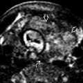
 tethered to each other (pseudosyndactyly) and to the thumb
  by a short band
  , which was best seen on real-time evaluation. Remember to use cine clips and high-resolution transducers for focused assessment of bands.
 . As an isolated finding, this would be of little consequence, but, in this case, the only other area involved was the umbilical cord, which was occluded by a band, resulting in intrauterine fetal demise.
 around the left calf with compromised flow in the posterior tibial artery
  . The anterior tibial artery
  is unaffected with good flow distal to the constriction. The infant had only extremity involvement and has done well with plastic surgery.
 from a constriction band. Another band is seen encircling the forearm
  .
.
Stay updated, free articles. Join our Telegram channel

Full access? Get Clinical Tree


