Aorto-Enteric Fistula
R. Brooke Jeffrey, MD
Key Facts
Terminology
Abnormal communication between aorta and gastrointestinal tract
Imaging
Best diagnostic clue: Inflammatory stranding and gas between abdominal aorta and 3rd part of duodenum following aneurysm repair
Location: Duodenum (80%); jejunum and ileum (10-15%); stomach and colon (5%)
Best imaging tool: CT 94% sensitive, 85% specific
Top Differential Diagnoses
Periaortitis
Retroperitoneal fibrosis
Postoperative changes
Postendovascular stent
Pathology
Primary etiology: Abdominal aortic aneurysms, infectious aortitis, penetrating peptic ulcer, tumor invasion, radiation therapy
Secondary etiology: Aortic reconstructive surgery most common
Clinical Issues
Very poor prognosis, up to 85% mortality
Percutaneous drainage of infected perigraft fluid may be initial treatment, followed by surgery
“Herald” GI bleeding, followed hours/days/weeks later by catastrophic hemorrhage
Diagnostic Checklist
Perigraft infection evidenced by ectopic gas or perigraft soft tissue raises suspicion of fistula
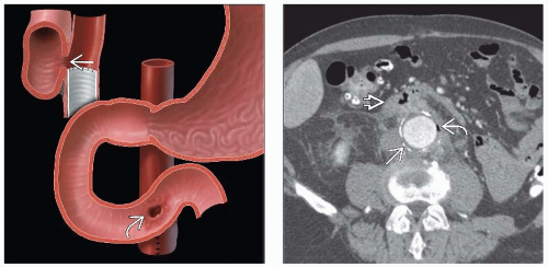 (Left) Graphic shows a fistula between the transverse duodenum & aorta
Get Clinical Tree app for offline access
 at the site of the graft-aortic suture line at the site of the graft-aortic suture line  . (Right) Axial CECT in a 70-year-old man presenting with fever and hematemesis months after abdominal aortic aneurysm repair shows the native, calcified aortic wall . (Right) Axial CECT in a 70-year-old man presenting with fever and hematemesis months after abdominal aortic aneurysm repair shows the native, calcified aortic wall  wrapped around a synthetic graft. A gas collection is noted between the graft and aortic wall wrapped around a synthetic graft. A gas collection is noted between the graft and aortic wall  , indicating infection or fistula. Note the soft tissue density surrounding the aorta and 3rd portion of duodenum , indicating infection or fistula. Note the soft tissue density surrounding the aorta and 3rd portion of duodenum
Related posts:Stay updated, free articles. Join our Telegram channel
Full access? Get Clinical Tree


|