Abstract
Balamuthia amoebic encephalitis (BAE) is a rare and often fatal central nervous system (CNS) infection caused by Balamuthia mandrillaris, a free-living amoeba typically found in soil and water. This organism can invade the brain directly, bypassing other organs, making early diagnosis particularly challenging. Symptoms often do not appear as distinctive early warning signs, and many patients do not experience noticeable skin lesions or systemic symptoms before neurological manifestations emerge. Balamuthia can enter the body through various routes, including the respiratory tract, skin, or gastrointestinal tract, eventually crossing the blood-brain barrier and causing aggressive encephalitis. The early symptoms of BAE are nonspecific, and the disease has an extremely high mortality rate. This report presents a 35-year-old male patient who died from Balamuthia amoebic encephalitis. The patient had a history of prolonged exposure to underground mines and consumed raw beef a week before the onset of symptoms. The infection is believed to have entered through the respiratory tract or gastrointestinal route. Diagnosis was primarily based on pathological findings, and the patient did not receive effective treatment due to delayed diagnosis, ultimately passing away approximately 2 months after the onset of symptoms. This case emphasizes the rarity and fatal nature of BAE, particularly when neurological symptoms are the first sign of infection without preceding systemic or dermatological manifestations. The report highlights the importance of considering Balamuthia mandrillaris infection in patients presenting with unexplained encephalitis and brain abscess, especially with a potential history of exposure to amoeba-contaminated environments.
Introduction
Balamuthia mandrillaris is a free-living amoeba found in soil and water that can cause Balamuthian amoebic encephalitis (BAE), a rare and fatal central nervous system (CNS) infection [ ]. The amoeba was first isolated from the brain of a baboon that died of meningoencephalitis [ ]. These amoebas usually enter the body through the skin, lungs, or gastrointestinal tract, ultimately crossing the blood-brain barrier and triggering amoebic encephalitis [ ]. BAE is extremely rare, with approximately 300 cases reported globally [ ]. Factors such as exposure to contaminated freshwater, soil, and immune suppression increase the risk of infection. Certain occupations, such as mining, plumbing, and agricultural work, also present a higher risk. The disease often begins insidiously, with nonspecific symptoms such as headache, fever, nausea, vomiting, seizures, confusion, vision loss, and speech difficulties [ ]. Due to its rapid progression and high fatality rate, early diagnosis is crucial to improve patient outcomes [ ].
Case reports
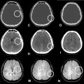
A 35-year-old male patient presented with a sudden onset of fever (38°C) on May 10, 2024, accompanied by abdominal pain, nausea, vomiting, and intermittent headache characterized by a pulsatile pain in the left eye socket and frontal region. The patient had not received any treatment, and there were no gastrointestinal symptoms such as diarrhea. On May 13, the patient developed right upper limb seizures with associated right-sided numbness, lasting for about 5 minutes before spontaneously resolving, with 5-6 episodes occurring daily. An MRI performed at a local hospital showed abnormal signals in the left parietal lobe, suggestive of a central nervous system infection. The patient was treated with anti-inflammatory medications, after which the seizures subsided, and his fever decreased to around 37°C. However, on May 15, the patient experienced progressive right-sided weakness, with difficulty using his right hand to hold chopsticks and walking difficulties in his right leg, accompanied by numbness in the right side of his body. On May 17, the patient was transferred to a higher-level hospital for further evaluation. MRI on May 22 revealed a round enhanced lesion in the left parietal lobe with associated edema, consistent with an infectious lesion. Despite treatment with antibiotics, corticosteroids, and medications to reduce intracranial pressure, the right-sided weakness persisted without significant improvement. Follow-up MRIs on June 4 and June 18 showed a reduction in the size of the lesion in the left parietal lobe ( Fig. 1 ) but the appearance of multiple new lesions in the right parietal lobe ( Fig. 2 ). On June 20, the patient developed left-sided numbness and weakness, unable to get out of bed, and had facial drooping. On June 24, due to worsening condition, the patient was transferred to our hospital’s neurology department for further management.


Stay updated, free articles. Join our Telegram channel

Full access? Get Clinical Tree


