KEY FACTS
Terminology
- •
Rare premalignant or malignant, unilocular or multilocular cystic tumor arising from biliary epithelium
- •
Synonyms: Hepatobiliary cystadenoma/carcinoma, biliary cystic tumor, biliary cystic neoplasm, mucinous cystic neoplasm of liver
Imaging
- •
Solitary, large, well-defined, multiloculated and multilobulated hepatic cyst
- ○
Thick, irregular wall and enhancing internal septations
- ○
May show biliary dilation from mass effect
- ○
- •
Biliary cystadenoma
- ○
Thin and smooth septa
- ○
May have fine calcifications and subtle mural nodularity (< 1 cm)
- ○
Absence of mural nodularity makes cystadenoma more likely
- ○
- •
Biliary cystadenocarcinoma more commonly associated with
- ○
Thick and irregular septa
- ○
Mural and septal nodularity (> 1 cm) and papillary projections
- ○
Coarse calcifications
- ○
Hemorrhagic internal fluid
- ○
- •
Location
- ○
Intrahepatic biliary ducts (83%), extrahepatic biliary ducts (13%), gallbladder (0.02%)
- ○
Top Differential Diagnoses
- •
Simple/complex/complicated hepatic cyst
- •
Hepatic abscess
- •
Echinococcal (hydatid) cyst
- •
Cystic metastases
Clinical Issues
- •
Primarily occurs in middle-aged white women
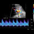
 with lobulated contour and multiple, irregular, vascularized septations
  .
.
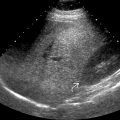
 . Most biliary cystadenomas are seen in middle-aged women.
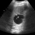
 and layering debris
  .
.


