KEY FACTS
Terminology
- •
Encapsulated collection of bile outside biliary tree
Imaging
- •
Ultrasound
- ○
Grayscale ultrasound
- –
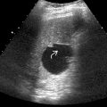
Focal collection of fluid within liver or close to biliary tree, or in gallbladder fossa in patients with recent cholecystectomy
- –
Round or oval in shape, usually unilocular, usually no discernible thin capsule
- –
Anechoic fluid content suggests fresh biloma
- –
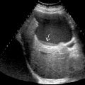
Debris or septa suggest infected biloma
- –
May see echogenic foci at periphery related to clips from recent surgery
- –
- ○
Color Doppler ultrasound
- –
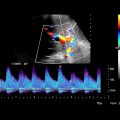
No vascularity within lesion
- –
For infected biloma, there may be increased vascularity in adjacent tissue
- –
- ○
- •
CECT: Well-defined or slightly irregular cystic lesion without identifiable wall
Top Differential Diagnoses
- •
Perihepatic collection/seroma/lymphocele
- •
Hepatic cyst
- •
Hepatic abscess
- •
Intrahepatic hematoma
Pathology
- •
Iatrogenic: Laparoscopic cholecystectomy, post liver transplantation, ERCP or other instrumentation of biliary tree, liver biopsy
- •
Post traumatic: Blunt trauma, motor vehicle accident
- •
Spontaneous rupture of bile duct
Scanning Tips
- •
Look for low-level echoes within biloma, which can indicate underlying infection
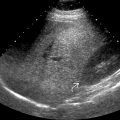
 after surgical removal of a liver mass. Low-level internal echoes
  suggest infected bile. Peripheral surgical suture with a ring-down artifact
  and clip with posterior shadowing
  are seen.
Stay updated, free articles. Join our Telegram channel

Full access? Get Clinical Tree


