Bladder Rupture
Cody J. Schwartz
CLINICAL HISTORY
50-year-old male with history of motor vehicle collision.
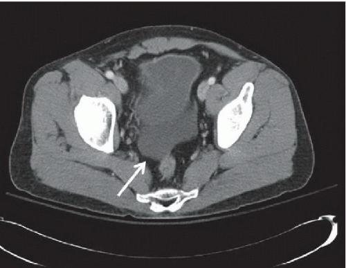 FIGURE 3A |
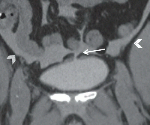 FIGURE 3B |
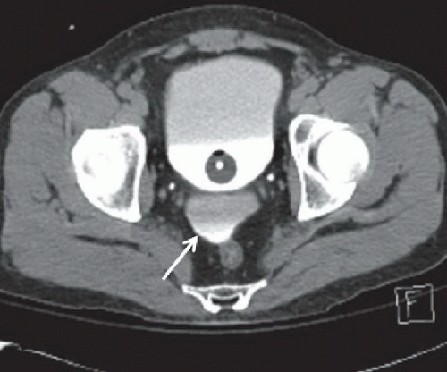 FIGURE 3C |
FINDINGS
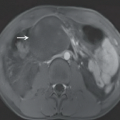
Figure 3A: Axial contrast-enhanced CT image of the pelvis demonstrates simple free fluid posterior to the bladder (arrow). Figure 3B: Axial CT image of the pelvis from a CT cystogram demonstrates hyperdense intraperitoneal fluid with layering extravasated intravesicular contrast in the posterior pelvis (arrow). A Foley catheter and layering contrast are identified in the bladder. Figure 3C: Coronal reformatted image of the pelvis from a CT cystogram showing hyperdense fluid in the bladder and in the intraperitoneal space. A defect is visible at the bladder dome, indicating the site of the bladder rupture with extravasation of intravesicular contrast from the bladder (arrow) into the peritoneal spaces along the mesentery, around bowel loops, and paracolic gutters (arrowheads).
Related posts:


Stay updated, free articles. Join our Telegram channel

Full access? Get Clinical Tree


