Abstract
An 80-year-old woman with a left breast mass was referred to our department. Mammography showed an oval mass, 2.5cm in size, with circumscribed margins in her left breast. Ultrasound showed an oval tumor with circumscribed margins, heterogenous internal echoes including numerous punctate hyperechoic foci, and posterior echo enhancement. Magnetic resonance imaging (MRI) of the tumor showed low and high signal intensity on T1-weighted images and on fat-suppressed T2-weighted images, respectively. Kinetic curve assessment of the tumor showed a fast and plateau pattern. After the pathological confirmation of malignant cells, the patient underwent mastectomy and sentinel node biopsy. Postoperative pathological study showed that atypical cells formed irregularly arranged papillary nests and grew in a medullary fashion accompanied by massive lymphocyte infiltration, leading to the diagnosis of invasive ductal carcinoma with medullary features (IDCMF). Immunostaining showed that the tumor had a triple negative phenotype and a high Ki-67 labelling index of 52%. In conclusion, breast diagnostic physicians should note that IDCsMF show a fast and plateau enhancement pattern on MRI kinetic curve assessment. Furthermore, the presence of punctate hyperechoic foci in the tumor can be useful in distinguishing IDCsMF from medullary breast carcinomas.
Introduction
Breast medullary carcinomas are rare breast malignancies, which have high nuclear grades, a triple negative phenotype, high proliferation potential, massive lymphocyte infiltration around the cancer cells, and no ductal structures. Despite their presumed aggressive characteristics, medullary carcinomas generally show favorable clinical outcomes [ , ].
Many researchers have reported that the abundant presence of tumor-infiltrating lymphocytes in the tumor contributes to the discrepancy between the dismal prognostic factors and the favorable clinical outcomes in medullary breast carcinomas [ ]. Breast cancers that resemble medullary carcinomas but have tubule forming structures are classified as invasive ductal carcinomas with medullary features (IDCsMF). Prominent lymphatic infiltration, however, is a frequent finding observed in both medullary carcinomas and IDCsMF.
We report a case of IDCMF which had characteristic image findings.
Case presentation
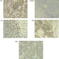
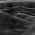
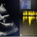
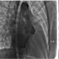
An 80-year-old woman with a family history of colon cancer and hepatocelular carcinoma in her 2 brothers was undergoing outpatient treatment at our hospital for primary biliary cholangitis. The patient also suffered from hypertension, dyslipidemia, and subclinical thyroid dysfunction, but noticed a painless mass in her left breast, leading to the consultation to our department. Mammography showed an oval mass, 2.5cm in size, with circumscribed margins in the upper outer quadrant of her left breast ( Fig. 1 ). Ultrasound showed that an oval tumor had circumscribed margins, heterogenous internal echoes including numerous punctate hyperechoic foci, and posterior echo enhancement ( Fig. 2 ). Magnetic resonance imaging (MRI) of the tumor showed low and high signal intensity on T1-weighted images and on fat-suppressed T2-weighted images, respectively. Kinetic curve assessment of the tumor showed a fast and plateau pattern. Enhanced areas, however, differed slightly between the early and late phase images ( Fig. 3 ). Under the tentative diagnosis of breast cancer, the patient underwent core needle biopsy of the breast mass. Pathological study showed nuclear grade 3 atypical cells growing in solid, trabecular, and papillary fashions with prominent inflammatory cell infiltration around the cancer cells. Immunostaining showed that the tumor was negative for estrogen receptor, progesterone receptor and human epidermal growth factor receptor type 2, and had a high Ki-67 labelling index of 45%. Due to no suspicious lymph nodes on palpation and ultrasound evaluation, the patient underwent mastectomy and sentinel node biopsy, resulting in no sentinel node metastasis on frozen section. Postoperative pathological study showed that atypical cells formed irregularly arranged papillary nests and grew in a medullary fashion accompanied by numerous lymphocyte infiltration. Immunostaining showed that the tumor had a triple negative phenotype and a higher Ki-67 labelling index of 52% than that observed in the core needle biopsy specimen ( Fig. 4 ). The patient was discharged in 7 days after operation and is scheduled to be followed-up without any adjuvant therapy due to her old age.

Related posts:
 Ovarian metastasis from lobular breast carcinoma: A case report with review of literature
 Invasive lobular carcinoma with metastasis to the pectoralis muscle
 Early onset development of hypertrophic cardiomyopathy in less than 1 year in a patient with familial Friedrich’s ataxia: Case report
 Radiologic evaluation of a giant aneurysmal left circumflex coronary artery presenting with palpitation: A Cas of successful diagnosis
 Giant ascending aortic aneurysm: A rare case report
 A rare and life-threatening case of spontaneous hemopneumothorax presenting with severe hemodynamic instability
Stay updated, free articles. Join our Telegram channel

Full access? Get Clinical Tree


