KEY FACTS
Terminology
- •
Budd-Chiari syndrome: Hepatic venous outflow obstruction
- •
Global or segmental obstruction of hepatic venous outflow or inferior vena cava (IVC)
Imaging
- •
Ultrasound acute phase
- ○
Absent or restricted flow, possible thrombosis in hepatic veins (HVs)/IVC
- ○
Intrahepatic collateralization, bicolored HVs: Flow in opposite direction in HV branches with common trunk
- ○
Reduced velocity, continuous flow in portal vein, possibly hepatofugal flow
- ○
- •
Ultrasound chronic phase
- ○
Hypertrophy of caudate lobe and unaffected segments, atrophy of involved segments, large regenerative nodules
- ○
Stenotic or occluded HVs/IVC
- ○
Intrahepatic &/or extrahepatic collateralization
- ○
- •
CECT: Flip-flop enhancement pattern
- ○
Early enhancement of caudate lobe and central portion around IVC, decreased peripheral liver enhancement
- ○
Later decreased enhancement centrally and increased enhancement peripherally
- ○
Large regenerated nodules, hypertrophic caudate lobe
- ○
Top Differential Diagnoses
- •
Liver cirrhosis
- •
Portal vein thrombosis
- •
Acute, severe passive venous congestion
- •
Acute hepatitis
Diagnostic Checklist
- •
Imaging interpretation pearls
- ○
Narrowed or obliterated HVs/IVC
- ○
Bicolored HVs due to intrahepatic collateralization on color Doppler ultrasound
- ○
Scanning Tips
- •
Use slow flow settings or B-flow imaging to show lack of flow in thrombosed hepatic veins
- •
May see bicolored HVs due to intrahepatic collateralization on color Doppler ultrasound
- •
Cine hepatic vein with grayscale (without moving transducer) to demonstrate flow (or confirm lack of flow) that is not detectable with color Doppler
- •
Thrombosed HVs may be barely discernible or cord-like, requiring extra attention to locate on grayscale
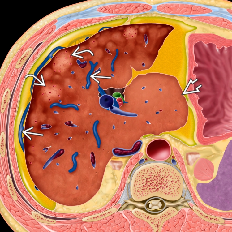
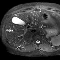
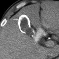
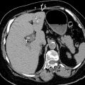
 , heterogeneous hepatic parenchyma due to centrilobular necrosis, and hypervascular regenerative nodules
  . Note the sparing of the caudate lobe with hypertrophy
  as well as the thrombosed inferior vena cava (IVC).