Cauda Equina Syndrome Secondary to Lumbar Disk Extrusion
Benjamin Y. Huang
CLINICAL HISTORY
79-year-old female presenting with back pain, lower extremity weakness, and urinary retention.
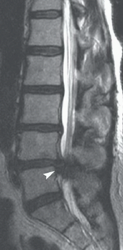 FIGURE 17A |
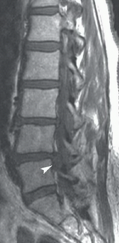 FIGURE 17B |
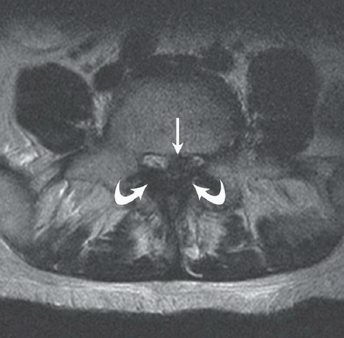 FIGURE 17C |
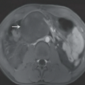
FINDINGS
Figures 17A and 17B: Sagittal T2-weighted (Fig. 17A) and T1-weighted (Fig. 17B) MR images through the lumbar spine demonstrating a globular, extradural soft tissue mass (arrowheads) posterior to the L4-L5 disk space, which appears contiguous with the intervertebral disk and of the same signal intensity as the disk on both pulse sequences. The mass extends slightly inferiorly relative to the disk level and completely obliterates the lumen of the spinal canal. Figure 17C: Axial T2WI just below the L4-L5 disk level again demonstrates the lesion (arrow), which on axial imaging has an ovoid shape and which, in combination with significant thickening of the ligamentum flavum (curved arrows), completely effaces the spinal canal. Note the complete absence of any visible CSF.
DIFFERENTIAL DIAGNOSIS
In this case, there are, clearly, findings worrisome for a spinal block at the L4-L5 level, and the differential becomes one of what is causing the stenosis. The primary diagnostic consideration in this case is an extruded (herniated) intervertebral disk fragment. When a disk herniation is suspected, efforts should be made to determine whether the disk fragment has become separated from its parent disk (sequestered disk). Alternative considerations for extradural mass lesions in the lumbar spine include a schwannoma, metastasis, epidural abscess, or epidural hematoma. Schwannomas generally arise from the spinal nerve roots and are therefore usually located more laterally in the neural foramina. Metastases usually spread into the epidural space from the adjacent vertebral bodies. In this case, the nearby L4 and L5 vertebrae demonstrate normal marrow signal. Epidural abscesses usually develop in the setting diskitis or septic arthritis. In this case, there are no findings to suggest an infectious etiology. Finally, epidural hematomas tend to be more longitudinally extensive, although they can be focal when associated with a fracture or acute disk herniation.
Related posts:


Stay updated, free articles. Join our Telegram channel

Full access? Get Clinical Tree


