Cerebellar Overview
Lubdha M. Shah, MD
IMAGING ANATOMY
Overview
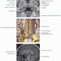
Cerebellum is composed of cortical gray matter surrounding subcortical white matter
Primary fissure divides anterior & posterior lobes
10 lobules at midline, 8 lobules in each hemisphere
Anterior lobe (lobules I-V); posterior lobe (lobules VI-IX); flocculonodular lobe (lobule X)
On ventral surface, posterolateral fissure separates posterior lobe from flocculonodular lobe
2 flocculi connected to midline nodulus, inferior part of vermis
Sagittal zones: Vermis, intermediate zone, lateral hemispheres
Arbor vitae: White matter carrying motor & sensory information to & from cerebellum
Deep gray nuclei (medial to lateral): Cerebellar output
Fastigial nucleus: Input from vermis & from
cerebellar afferents that carry vestibular, proximal somatosensory, auditory, & visual info
Projects to vestibular nuclei & reticular formation
Interposed nuclei (globose & emboliform nuclei): Input from intermediate zone & from cerebellar afferents, carrying spinal, proximal somatosensory, auditory, & visual info
Project to contralateral red nucleus
Dentate nucleus: Input from lateral hemisphere & from cerebellar afferents carrying information from cerebral cortex (via pontine nuclei)
Project to contralateral red nucleus & ventrolateral thalamic nucleus
Vestibular nuclei: In medulla
Functionally equivalent to cerebellar nuclei because of same connectivity patterns
Input from flocculonodular lobe & vestibular labyrinth
Project to various motor nuclei & originate the vestibulospinal tracts
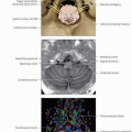
Cerebellar peduncles (CP)
Superior CP: Efferents from cerebellum
Middle CP: Afferents to cerebellum
Inferior CP: Afferents to cerebellum
ANATOMY IMAGING ISSUES
Imaging Recommendations
Increased spatial resolution of ultrahigh field enables visualization of deep cerebellar nuclei & cerebello-cortical sublayers
Diffusion tensor tractography reconstructs afferent & efferent cerebellar pathways
Diffusion spectrum imaging able to resolve complex distributions of intravoxel fiber orientations & fiber crossings within neural structures
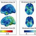
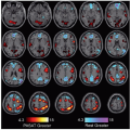
Resting-state functional connectivity individualizes prewired, parallel close-looped sensorimotor, cognitive, & affective networks passing through cerebellum
Activity in sensorimotor regions correlates with contralateral cerebellar anterior lobe & lobule VIII
Activity in prefrontal, posterior parietal, & superior & middle temporal association areas, as well as cingulate gyrus & retrosplenial cortex, correlates with activity in lobules VI and VII
fMRI studies
Stay updated, free articles. Join our Telegram channel

Full access? Get Clinical Tree


