TERMINOLOGY
Synonyms
- •
Internal jugular chain (IJC): Deep cervical chain
- •
Spinal accessory chain (SAC): Posterior triangle chain
- •
Transverse cervical chain: Supraclavicular chain
- •
Anterior cervical chain: Prelaryngeal, pretracheal, paratracheal nodes
- •
Paratracheal node: Recurrent laryngeal node
Definitions
- •
Jugulodigastric node: “Sentinel” (highest) node, found at apex of IJC at angle of mandible
- •
Virchow node: “Signal” node, lowest node of deep cervical chain
- •
Troisier node: Most medial node of transverse cervical chain
- •
Omohyoid node: Deep cervical chain node superior to omohyoid as it crosses jugular vein
- •
Delphian node: Pretracheal node
IMAGING ANATOMY
Overview
- •
In normal adult neck, may be up to 300 lymph nodes
- ○
Internal structures: Capsule, cortex, medulla, hilum
- ○
- •
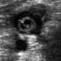
US appearances of normal cervical lymph node
- ○
Small, oval/reniform shape with well-defined margin
- ○
Homogeneous, hypoechoic cortex with echogenic fatty hilum
- ○
Hilar vascularity on color/power Doppler examination
- ○
- •
Imaging-based nodal classification
- ○
Level I: Submental and submandibular nodes
- –
Level IA: Submental nodes: Found between anterior bellies of digastric muscles
- –
Level IB: Submandibular nodes: Found around submandibular glands in submandibular space
- –
- ○
Level II: Upper IJC nodes: From posterior belly of digastric muscle to hyoid bone
- –
Level IIA: Level II node anterior, medial, lateral, or posterior to IJV; if posterior to IJV, node must be inseparable from IJV; contains jugulodigastric nodal group
- –
Level IIB: Level II node posterior to IJV with fat plane visible between node and IJV
- –
- ○
Level III: Mid IJC nodes
- –
From hyoid bone to inferior margin of cricoid cartilage
- –
- ○
Level IV: Lower IJC nodes
- –
From inferior cricoid margin to clavicle
- –
- ○
Level V: Nodes of posterior cervical space/spinal accessory chain
- –
SAC nodes lie posterior to back margin of sternocleidomastoid muscle
- –
Level VA: Upper SAC nodes from skull base to bottom of cricoid cartilage
- –
Level VB: Lower SAC nodes from cricoid to clavicle
- –
- ○
Level VI: Nodes of visceral space
- –
Found from hyoid bone above to top of manubrium below
- –
Midline group of cervical lymph nodes
- –
Includes prelaryngeal, pretracheal, and paratracheal subgroups
- –
- ○
Level VII: Superior mediastinal nodes
- –
Between carotid arteries from top of manubrium above to innominate vein below
- –
- ○
- •
Other nodal groups not included in standard imaging-based nodal classification
- ○
Parotid nodal group: Intraglandular or extraglandular
- ○
Retropharyngeal (RPS) nodal group: Medial RPS nodes and lateral RPS nodes (Rouvière node)
- ○
Facial nodal group
- ○
ANATOMY IMAGING ISSUES
Imaging Approaches
- •
Nodal metastases from primary tumors are site specific; therefore, it is critical to understand usual patterns of lymphatic spread
- •
Equivocal nodes outside usual pattern less suspicious
- •
Likely location of primary tumor can be suspected in patients presenting with nodal mass
- •
Nodal disease outside usual pattern may suggest aggressive tumor or prompt search for 2nd primary
Imaging Pitfalls
- •
RPS nodes and superior mediastinal nodes cannot be assessed by US
Key Concepts
- •
Useful US features suspicious of malignancy
- ○
Shape: Round, long:short axis ratio < 2
- ○
Loss of echogenic hilum
- ○
Presence of intranodal necrosis (cystic/coagulation)
- ○
Presence of extracapsular spread: Ill-defined margin
- ○
Peripheral/subcapsular flow on color/power Doppler ultrasound
- ○
Increased intranodal intravascular resistance: Resistive index (RI) > 0.8, pulsatility index (PI) > 1.6
- ○
Internal architecture: Punctate calcifications in metastatic node from papillary thyroid carcinoma, reticulated/pseudocystic appearance of lymphomatous node
- ○
- •
No single finding sensitive or specific enough; these signs should be used in combination
- •
Fine-needle aspiration biopsy helps to improve diagnostic accuracy
- •
Tuberculous nodes mimic metastatic nodes
- ○
Differentiating features: Intranodal necrosis, nodal matting, soft tissue edema and displaced hilar vascularity/avascularity, calcification (post treatment)
- ○
CLINICAL IMPLICATIONS
Clinical Importance
- •
Presence of malignant SCCa nodes on staging associated with 50% ↓ in long-term survival
- ○
If extranodal spread present, further 50% ↓
- ○
- •
Location of metastatic nodes in neck may help predict site of primary tumor
- ○
RPS and posterior triangle nodes seen in nasopharyngeal carcinoma, and lower cervical nodes in lung cancer
- ○
When Virchow node found on imaging without upper neck nodes, primary not in neck, and whole-body imaging warranted
- ○
LYMPH NODE GROUPS