Abstract
Sarcoidosis is a systemic inflammatory disease characterized by the formation of noncaseating granulomas. While commonly affecting the lungs and lymph nodes, nasosinusal involvement is rare, accounting for less than 1% of cases. When the nasosinusal region is affected, clinical manifestations may include persistent nasal obstruction, epistaxis, anosmia, and facial. Diagnosis relies on imaging studies, which may reveal mucosal thickening or destructive lesions, and histopathological confirmation through biopsy. Management typically involves systemic corticosteroids, with immunosuppressive agents reserved for refractory cases. This uncommon localization can mimic other inflammatory or infectious conditions, underscoring the importance of accurate diagnosis for appropriate treatment. We report the case of a 55-year-old female patient with a history of chronic rhinosinusitis persisting for 2 years despite conventional treatment, who presented to our facility with a clinical picture of chronic nasal obstruction rhinorrhea and anosmia. Clinical, laboratory, and imaging investigations confirmed the diagnosis of nasosinusal sarcoidosis. Given the rarity of this condition, maintaining a high index of clinical suspicion is crucial to prevent misdiagnosis and delays in initiating appropriate treatment, which is essential for optimizing the patient’s outcome.
Introduction
Sarcoidosis is a systemic inflammatory disease of unknown etiology, characterized by the formation of noncaseating granulomas affecting multiple organ systems. Among its extrathoracic manifestations, otorhinolaryngological (ENT) involvement is observed in 5%-15% of cases and may represent the initial presentation of the disease. Sinonasal sarcoidosis, although rare, occurs in 1%-4% of sarcoidosis cases and is a well-recognized yet chronic and challenging form of the disease [ ].
Typical symptoms include chronic crusting rhinitis, nasal obstruction, and anosmia, with severe cases leading to structural and functional complications. This complex condition highlights the diagnostic and therapeutic challenges posed by ENT sarcoidosis, often requiring a multidisciplinary approach that combines pharmacological treatments and targeted surgical interventions [ ].
The management of nasosinusoidal sarcoidosis involves the use of intranasal corticosteroids to reduce local inflammation. In cases of more severe symptoms, systemic corticosteroids may be required. For refractory disease, immunosuppressive agents such as methotrexate or biological therapies can be considered. Regular monitoring is essential to adjust treatment and prevent complications [ ].
Case presentation
A 55-year-old female patient presented with a 2-year history of chronic rhinosinusitis, having undergone several cycles of nasal corticosteroid and antibiotic treatments. She came to our clinic with persistent bilateral nasal obstruction, purulent rhinorrhea, and anosmia. Nasal endoscopy revealed mucosal inflammation, including edema of the inferior turbinates, presence of synechiae, and the formation of crusts. The rest of the ENT and general examination was unremarkable.
A laboratory work-up showed lymphocytosis (5500/mm³) (Normal range 1000-4000/mm³) and hypercalcemia (18.5 mg/dL) (Normal range 8.5-10.5mg/dl),with no other abnormalities.
Nasosinus CT scans demonstrated irregular hypertrophy of the nasal mucosa and bilateral enlargement of the inferior turbinates, as well as a frame-like mucosal thickening in the right maxillary sinus ( Fig. 1 ).

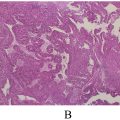
During a multidisciplinary consultation, taking into account clinical findings, laboratory results, and imaging studies, and despite the lack of symptomatic improvement with medical treatment, a nasal mucosal biopsy was performed, revealing an epithelioid giant cell granuloma without caseating necrosis ( Figs. 2 , 3 ). A diagnosis of isolated nasosinus sarcoidosis was confirmed.


The urgent management plan included systemic oral corticosteroids at a dosage of 1 mg/kg/day for 3 months, supplemented with localized corticosteroid therapy and nasal irrigation for 6 months. Clinical progress was notably favorable, with follow-up imaging showing a significant reduction in the size of the turbinates and substantial improvement in laboratory parameters. By the first month postdiagnosis, there was normalization of lymphocyte count and marked resolution of hypercalcemia, indicating resolution of the infection and attenuation of the systemic inflammatory response.
Discussion
Sarcoidosis is a multisystemic inflammatory disease characterized by the formation of noncaseating granulomas. Although sinonasal involvement is rare, it represents a significant diagnostic and therapeutic challenge due to its nonspecific symptoms and potential overlap with other granulomatous diseases. Sinonasal sarcoidosis accounts for approximately 0.6%-4% of sarcoidosis cases, with variability depending on study populations and diagnostic criteria [ ].
Sarcoidosis predominantly affects young to middle-aged adults, with a higher prevalence among women and African Americans. Sinonasal involvement is less common but may occur as an isolated manifestation or in conjunction with systemic disease. The underdiagnosis of sinonasal sarcoidosis is likely due to its subtle presentation and its rarity compared to pulmonary manifestations [ ].
The pathogenesis of sarcoidosis involves a dysregulated immune response triggered by an unidentified antigen [ ]. The key immunological process includes:
- •
Activation of antigen-presenting cells and recruitment of CD4+ T cells.
- •
Production of proinflammatory cytokines, including tumor necrosis factor-alpha (TNF-α), interleukin-12 (IL-12), and interferon-gamma (IFN-γ), which perpetuate granuloma formation.
- •
Genetic predisposition involving specific HLA alleles, such as HLA-DRB1, contributes to susceptibility and phenotype variability.
Environmental exposures, such as bioaerosols and microbial agents, have been proposed as potential triggers [ ].
Sinonasal sarcoidosis is a rare and chronic manifestation of sarcoidosis, affecting 1%-4% of sarcoidosis patients. Its diagnosis is based on a combination of clinical, radiological, and histopathological findings [ ].
Patients commonly present with symptoms such as chronic crusting rhinitis (70%-90%), unilateral or bilateral nasal obstruction (80%-90%), anosmia (approximately 70%), and epistaxis (20%). Clinical examination may reveal hypertrophy and a purplish discoloration of the nasal mucosa, often accompanied by granulomatous changes on the septum or inferior turbinates. Nasal polyps are observed in 35%-40% of cases, along with turbinoseptal synechiae (11%-13%), septal perforation (11%), and external nasal deformities (10%) [ ].
Computed tomography and magnetic resonance imaging are essential tools for identifying nodular lesions of the septum or inferior turbinates, mucosal thickening, and complete or partial opacification of the ethmoidal, maxillary, or sphenoidal sinuses. These radiological changes are present in 50%-85% of SNS cases [ ].
The diagnosis is confirmed by identifying noncaseating epithelioid granulomas in biopsy specimens of affected tissues, typically obtained from the nasal mucosa. It is crucial to exclude other causes of granulomatous diseases, such as tuberculosis, atypical mycobacteria, or granulomatosis with polyangitis [ ].
Conditions that mimic SNS include nasal tuberculosis, aspergillosis, actinomycosis, granulomatosis with polyangiitis, Churg-Strauss syndrome, and rare sinonasal manifestations of Crohn’s disease [ ].
This multi-faceted diagnostic approach ensures accurate identification of sinonasal sarcoidosis and provides the foundation for appropriate therapeutic management [ ].
The management and progression of sinonasal sarcoidosis require a multidisciplinary approach. This rare condition, challenging to diagnose due to its nonspecific clinical presentations, warrants high diagnostic suspicion in cases of chronic rhinosinusitis unresponsive to standard treatment. Therapy primarily relies on corticosteroids, administered either locally (intranasal corticosteroids) or systemically, depending on the disease’s severity. Systemic corticosteroids often yield incomplete responses, necessitating adjunctive immunosuppressive therapies such as methotrexate, azathioprine, or TNF-α inhibitors for refractory cases or when steroid-related adverse effects are substantial [ ].
In severe or destructive forms, surgical interventions, including endoscopic sinus surgery or nasal reconstruction, may be considered. However, these procedures are reserved for cases unresponsive to medical treatments or presenting with mechanical complications. Clinical outcomes remain variable: while some patients achieve spontaneous remission or significant improvement under treatment, others experience chronic progression with risks of nasal fibrosis and significant functional impairment [ ].
Recent data emphasize the importance of a tailored approach, integrating corticosteroids, immunosuppressants, and, in selected cases, biologic agents such as infliximab. These therapeutic strategies, combined with close clinical and radiologic monitoring, improve symptoms in most cases, although a notable subset may remain refractory to treatment [ ].
Conclusion
Sinonasal sarcoidosis, though rare, is an important differential diagnosis in patients presenting with chronic sinonasal symptoms unresponsive to standard therapies. A multidisciplinary approach involving otolaryngologists, pulmonologists, and immunologists is essential for optimal management. Further research is needed to establish standardized treatment protocols and explore targeted therapies for this challenging condition.
Patient consent
The patient has provided informed consent for the publication of this case report.
Competing Interests: The authors declare no conflicts of interest.
Acknowledgments: We extend our gratitude to the otorhinolaryngology department team at the university hospital for their management and availability. This study did not receive any specific funding from public, commercial, or nonprofit funding agencies.
References
Stay updated, free articles. Join our Telegram channel

Full access? Get Clinical Tree


