KEY FACTS
Terminology
- •
Cleft lip (CL) occurs ± cleft palate (CP)
- ○
> 80% of fetuses with CL also have CP
- ○
- •
Variable classifications, best to be descriptive about defects
- ○
Unilateral (right or left), bilateral, midline
- ○
Complete CL: Defect extends to nose (nares)
- ○
Incomplete CL: Cleft does not extend to nares
- ○
- •
Primary palate: Front of palate (alveolar ridge)
- •
Secondary palate: Back of palate (soft + hard palate)
Imaging
- •
Unilateral CL + CP is most common type
- ○
CL extends to nose and flattens nares
- ○
Seen best on standard coronal nose-lip view
- ○
- •
Bilateral CL/CP
- ○
Alveolar ridge becomes mass-like and bulges forward
- ○
Profile view best to see premaxillary protrusion
- ○
- •
Midline CL/CP: Central lip and palate defect
- ○
Flat midface and nose
- ○
- •
Isolated CP without CL is highly associated with small chin
- ○
Palate defect is deep (posterior to alveolar ridge)
- ○
- •
3D ultrasound helpful for making specific diagnosis
- ○
Helps family and maxillofacial team prepare
- ○
- •
Associations: Aneuploidy, brain, and cardiac anomalies
- ○
Trisomy 13, trisomy 18, > 200 syndromes
- ○
More likely with midline and bilateral CL/CP
- ○
Scanning Tips
- •
Best to obtain 3D volume from profile view
- ○
Use soft tissue post processing to determine if CL extends to nares and their appearance (often flattened)
- ○
Use axial plane and bone postprocessing coronal images to best evaluate palate defect
- ○
- •
When chin is small, look for fluid extending from mouth to nasal cavity posteriorly (soft tissue palate defect)
- •
Scan carefully for other anomalies, especially brain and heart

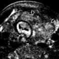
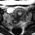
 . The flattened naris
  is seen best with 3D. This fetus also had CP. Treatment includes multiple surgeries and presurgical nasal alveolar molding.
 (and beyond).


