Clinical and Imaging Features of the Arthritides and Arthropathies
CLINICAL FEATURES
The accurate diagnosis of specific arthritis or arthropathy depends on many factors; however, the most important is to understand the patterns of symptoms and the mechanism of disease.
The clinical manifestations and laboratory data, in conjunction with the imaging findings, are of significant help in making the diagnosis of a specific arthritic process. The various arthritides, for example, have different frequencies of occurrence between the genders. Rheumatoid arthritis is much more common in females, and erosive osteoarthritis is seen almost exclusively in middle-aged women. Psoriatic arthritis, reactive arthritis (formerly known as a Reiter syndrome), and gouty arthritis are more common in males. Clinical symptoms are of further assistance. Patients with reactive arthritis, for example, usually present with urethritis, conjunctivitis, and mucocutaneous lesions, and those with psoriatic arthritis may present with swelling of a single finger, the so-called sausage digit (Fig. 3.1; see also Fig. 1.4G) as well as changes in the skin and fingernails (Fig. 3.2; see also Fig. 1.4A-F). Patients with gouty arthritis may exhibit soft tissue masses, representing chronic tophi, on the dorsal aspect of the hands or feet (see Fig. 1.5). Patients with inflammatory arthritis commonly exhibit swollen joints and alignment deformities (Fig. 3.3; see also Figs. 1.2 and 1.3).
Laboratory data are also essential. Gouty arthropathy, for instance, is associated with elevated serum uric acid concentrations, and examination of synovial fluid reveals monosodium urate crystals (see Fig. 1.11). The synovial fluid of patients with calcium pyrophosphate dihydrate (CPPD) crystal deposition disease contains calcium pyrophosphate crystals (see Figs. 1.9 and 1.10). Findings of ragocytes with cytoplasmatic inclusions of ingested aggregated IgG immunoglobulin within the synovial fluid of patients with rheumatoid arthritis can be a significant diagnostic feature (see Fig. 1.7). The detection of autoantibodies is another important aid in the diagnostic workup. Rheumatoid factor (RF) is a typical finding in rheumatoid arthritis. Patients lacking the specific
antibodies represented by RF are said to have “seronegative” arthritis. Anti-cyclic citrullinated peptide antibodies are also specific for RA. Patients with lupus arthritis have a positive lupus erythematosus cell test. The presence of a positive ANA is one of the most important abnormalities detected in patients with SLE, although it may be positive in some other autoimmune conditions. The presence of anti-dsDNA, anti-Sm, and anti-RNP antibodies is another highly specific factor for the diagnosis. Lastly, identification of the antigens of the major histocompatibility complex, particularly human leukocyte-associated antigens HLA-B27 and HLA-DR4, has in recent years become the crucial tests in the diagnosis of arthritic disease. It has been reported that 95% of patients with ankylosing spondylitis, 86% of patients with reactive arthritis, and 60% of patients with psoriatic arthropathy test positively for antigen HLA-B27, whereas a majority of those with rheumatoid arthritis exhibit the HLA-DR4 antigen. This is helpful in differentiating certain types of arthritides, as well as distinguishing psoriatic arthritis from rheumatoid arthritis in cases in which the radiographic presentation of these conditions may be very similar.
antibodies represented by RF are said to have “seronegative” arthritis. Anti-cyclic citrullinated peptide antibodies are also specific for RA. Patients with lupus arthritis have a positive lupus erythematosus cell test. The presence of a positive ANA is one of the most important abnormalities detected in patients with SLE, although it may be positive in some other autoimmune conditions. The presence of anti-dsDNA, anti-Sm, and anti-RNP antibodies is another highly specific factor for the diagnosis. Lastly, identification of the antigens of the major histocompatibility complex, particularly human leukocyte-associated antigens HLA-B27 and HLA-DR4, has in recent years become the crucial tests in the diagnosis of arthritic disease. It has been reported that 95% of patients with ankylosing spondylitis, 86% of patients with reactive arthritis, and 60% of patients with psoriatic arthropathy test positively for antigen HLA-B27, whereas a majority of those with rheumatoid arthritis exhibit the HLA-DR4 antigen. This is helpful in differentiating certain types of arthritides, as well as distinguishing psoriatic arthritis from rheumatoid arthritis in cases in which the radiographic presentation of these conditions may be very similar.
 Figure 3.1 ▪ Psoriatic arthritis—sausage digits. Diffuse swelling of the index and ring fingers represents dactylitis. Observe also typical for psoriasis skin changes. |
 Figure 3.2 ▪ Psoriatic arthritis—nail abnormalities. A: Pitting. B: Leukonychia. C: Splinter hemorrhages. D: Subungual hyperkeratosis. |
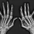
 Figure 3.3 ▪ Rheumatoid arthritis—joints malalignments. Ulnar deviation in the metacarpophalangeal joints is accompanied by swelling. Observe also hyperextension of the thumb, known as a “hitchhiker’s thumb” deformity. (From Bullough PG. Atlas of Orthopedic Pathology with Clinical and Radiologic Correlation. 2nd ed. New York: Gower Medical Publishing; 1992, Fig. 11.2.) |
IMAGING FEATURES
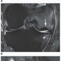
The true or diarthrodial joint consists of cartilage covering the articular ends of the bones forming the joint (see Fig. 1.18A); the articular capsule, which is reinforced by ligamentous structures; and the joint space, which is lined with synovial membrane and filled with synovial fluid (Fig. 3.4). Because of its physicochemical constitution, articular cartilage absorbs only a minimal amount of x-rays, thus appearing radiolucent on a radiographic film. The radiolucent articular cartilage, together with the joint cavity filled with synovial fluid, creates the so-called radiographic joint space.
 Figure 3.4 ▪ The constituent structures of a synovial (diarthrodial) joint. |
The abnormality of the joint in arthritis usually consists of destruction of the articular cartilage, which appears on radiographs as a narrowing of the radiographic joint space, usually accompanied by subchondral erosion; narrowing of the joint is the cardinal sign of arthritis (Fig. 3.5). It should be kept in mind, however, that in some arthritic processes, the joint space may not become narrow, appearing instead slightly expanded. This happens, for example, in the early stages of some arthritides, when joint effusion and ligamentous laxity cause distention of the joint with fluid, but the articular cartilage has not yet been destroyed. It may also be seen in rare instances when granulation pannus erodes the subchondral bone without destroying the articular cartilage (Fig. 3.6).
Other radiographic signs specific to different types of arthritis include periarticular soft tissue swelling, periarticular osteoporosis, and, in the more advanced stages of some arthritides, complete destruction of the joint with subluxation or dislocation and ankylosis (joint fusion) (Fig. 3.7).
The radiographic presentation of arthritis depends on the type and stage of the disease, as well as the site of the original insult characteristic for the various forms of arthritis or arthropathy (Fig. 3.8)—whether it is the articular cartilage, as in osteoarthritis (Fig. 3.9); the synovial membrane, as in inflammatory arthritis (Fig. 3.10); the synovial membrane, subchondral bone, and periarticular soft tissues, as in infectious arthritis (Fig. 3.11); or the synovial membrane, articular cartilage, subchondral bone, and periarticular soft tissues, as in some metabolic arthropathies (Fig. 3.12).
 Figure 3.5 ▪ Narrowing of the joint space. The cardinal sign of an arthritic process is narrowing of the radiographic joint space. Thinning of the articular cartilage reduces the space mechanically. |
 Figure 3.6 ▪ A and B: Variations in the width of the joint space. |
The radiographic diagnosis of arthritis, as Resnick observed, is based on the evaluation of two fundamental parameters: the morphology of the articular lesion and its distribution in the skeleton. If these findings are combined with the history, physical examination, and relevant laboratory data in a given case, then the accuracy of the diagnosis is markedly improved.
 Figure 3.7 ▪ Radiographic features of various arthritides. Summary representation of radiographic features seen in the arthritides. Not all of these features are seen in every type of arthritis. |
Morphology of the Articular Lesion
The various arthritides and arthropathies exhibit morphologically distinct features, as observed radiographically in the large (Fig. 3.13) and small (Fig. 3.14) joints. In the degenerative form of the disease known as osteoarthritis (osteoarthrosis), thinning of the articular cartilage results in localized narrowing of the joint space; there is also
subchondral sclerosis and osteophyte and cyst formation, but generally osteoporosis is absent (Fig. 3.15). Erosive osteoarthritis is characterized by central articular erosions and marginal proliferation of bone assuming the so-called gull-wing deformity (Fig. 3.16). Inflammatory arthritides, such as rheumatoid arthritis, are characterized by a diffuse, usually multicompartmental narrowing of the joint space associated with marginal or central erosions, periarticular osteoporosis, and symmetric periarticular soft tissue swelling; subchondral sclerosis is minimal or absent,
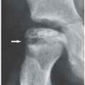
and formation of osteophytes is lacking (Fig. 3.17). In a metabolic arthropathy such as gout, well-defined osseous (articular or periarticular) erosions, displaying a so-called overhanging edge, are usually associated with preservation of part of the joint space and localized, asymmetric soft tissue nodules; osteophyte formation and osteoporosis are absent (Fig. 3.18). Infectious arthritis is characterized by the complete destruction of both articular ends of the bones forming the joint; all communicating joint compartments are invariably involved, with diffuse osteoporosis, joint effusion, and periarticular soft tissue swelling (Fig. 3.19). Neuropathic arthropathy is marked
by destruction of the articular surfaces, which leaves bony debris, and a substantial joint effusion; osteoporosis is usually lacking. Depending on the amount of destruction, varying degrees of joint instability are present (Fig. 3.20).
subchondral sclerosis and osteophyte and cyst formation, but generally osteoporosis is absent (Fig. 3.15). Erosive osteoarthritis is characterized by central articular erosions and marginal proliferation of bone assuming the so-called gull-wing deformity (Fig. 3.16). Inflammatory arthritides, such as rheumatoid arthritis, are characterized by a diffuse, usually multicompartmental narrowing of the joint space associated with marginal or central erosions, periarticular osteoporosis, and symmetric periarticular soft tissue swelling; subchondral sclerosis is minimal or absent,
and formation of osteophytes is lacking (Fig. 3.17). In a metabolic arthropathy such as gout, well-defined osseous (articular or periarticular) erosions, displaying a so-called overhanging edge, are usually associated with preservation of part of the joint space and localized, asymmetric soft tissue nodules; osteophyte formation and osteoporosis are absent (Fig. 3.18). Infectious arthritis is characterized by the complete destruction of both articular ends of the bones forming the joint; all communicating joint compartments are invariably involved, with diffuse osteoporosis, joint effusion, and periarticular soft tissue swelling (Fig. 3.19). Neuropathic arthropathy is marked
by destruction of the articular surfaces, which leaves bony debris, and a substantial joint effusion; osteoporosis is usually lacking. Depending on the amount of destruction, varying degrees of joint instability are present (Fig. 3.20).
 Figure 3.8 ▪ Target sites of various arthritides and arthropathies. |
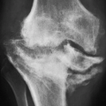
 Figure 3.9 ▪ Osteoarthritis. A 58-year-old woman presented with a history of pain in the left knee. A: Anteroposterior radiograph of the knee demonstrates narrowing of the medial femorotibial joint compartment and marginal osteophytes arising from both the medial and lateral femoral condyles—findings typical of osteoarthritis (degenerative joint disease). B: Lateral radiograph demonstrates, in addition, osteophytes at the anterior and posterior aspects of the articular end of the tibia, which are not appreciated on the anteroposterior projection. Involvement of the femoropatellar joint compartment and the presence of synovitis, represented by suprapatellar joint effusion, are also well demonstrated. C: Conventional radiograph of the hip demonstrates the typical morphologic changes seen in degenerative joint disease (osteoarthritis): focal narrowing of the joint space (here at the weight-bearing segment), subchondral sclerosis, cystlike lesions, and marginal osteophytes. Note the lack of osteoporosis. D: Anteroposterior radiograph of the right shoulder of a 58-year-old man shows the typical features of osteoarthritis, including joint space narrowing, subchondral sclerosis, and osteophyte formation.
Related posts:Stay updated, free articles. Join our Telegram channel
Full access? Get Clinical Tree
 Get Clinical Tree app for offline access

|