Abstract
Gallbladder lesion characterization remains a common diagnostic dilemma in abdominal imaging, particularly when differentiating between polyps and organized sludge. Findings on conventional gray-scale ultrasound may be equivocal, especially when typical imaging features such as mobility are absent. We present a case of a 65-year-old patient who presented with a concerning nonmobile 3.7 cm gallbladder lesion on initial ultrasound assessment. Due to the superior sensitivity of contrast-enhanced ultrasound (CEUS) for blood flow compared to other imaging modalities, CEUS was able to demonstrate complete absence of enhancement within the lesion, consistent with tumefactive sludge rather than solid tissue, and averting the need for additional cross-sectional imaging. Surgical pathology following cholecystectomy confirmed these findings. This case highlights the utility of CEUS as a valuable tool in gallbladder imaging, potentially reducing healthcare costs and expediting appropriate patient care while avoiding the risks associated with other contrast-enhanced imaging modalities.
Introduction
Gallbladder lesions represent a common diagnostic challenge in abdominal imaging, with the differentiation between neoplastic processes and benign findings having significant clinical implications [ ]. While conventional gray-scale ultrasound remains the primary imaging modality for gallbladder assessment, its ability to definitively characterize lesions can be limited, particularly when typical imaging features are absent or equivocal [ , ]. Traditionally, mobility with positional changes has served as a reliable indicator for confirming that a sludge ball is not a true soft tissue lesion; however, diagnostic uncertainty arises when sludge becomes organized and demonstrates mass-like features [ ].
Current imaging algorithms frequently suggest advancing to magnetic resonance imaging (MRI) for further characterization of indeterminate gallbladder lesions [ ]. This approach, while effective, often results in increased imaging costs, potential delays in care due to limited resources, and barriers for patients with MRI contraindications such as cardiac pacemakers [ ]. Furthermore, MRI (along with CT) carries risks associated with conventional contrast agents, particularly in patients with renal dysfunction [ ].
Contrast-enhanced ultrasound has emerged as a valuable problem-solving tool in abdominal imaging, utilizing an intravenously injected microbubble contrast agent to offer real-time assessment of tissue perfusion patterns without ionizing radiation or risk of nephrotoxicity [ ]. The use of ultrasound contrast agents, such as Definity (Perflutren lipid microsphere), allows for dynamic evaluation of lesion enhancement patterns, potentially obviating the need for more expensive cross-sectional imaging [ ]. While CEUS has gained widespread acceptance in European and Asian practice for various applications, including gallbladder assessment, it remains underutilized in North America [ ]. Recent literature suggests that CEUS may offer comparable diagnostic accuracy to MRI for gallbladder lesion characterization while providing advantages in terms of cost, availability, and patient safety [ , ].
Case report
A 65-year-old male patient with no known risk factors for gallbladder cancer was referred for ultrasound evaluation of gallbladder lesions initially detected on outside imaging. The imaging had been performed in 2016 to investigate right upper quadrant and epigastric pain. He had a past medical history of dyslipidemia, hypertension, gastroesophageal reflux disease (GERD), obstructive sleep apnea, and osteoarthritis. He was a former smoker with a 30 pack-year history, having quit 19 years prior. His surgical history included a prior appendectomy. The patient had no relevant family history.
At the initial presentation, the patient reported intermittent right upper quadrant and epigastric pain, prompting an initial abdominal ultrasound in January 2016. This study revealed hepatomegaly with hepatic steatosis, mild gallbladder wall thickening (3.8 mm) without evidence of cholelithiasis or acute cholecystitis. Laboratory investigations showed a total bilirubin of 9 µmol/L, AST of 37 U/L, ALT of 26 U/L, ALP of 53 U/L, GGT of 136 U/L, and a random glucose level of 7.2 mmol/L. His complete cardiac workup, performed concurrently for unrelated chest pain, was unremarkable, including negative troponins, normal ECG, and a normal stress test.
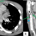
Conventional ultrasound performed at our hospital 2 months later revealed a 3.7 cm intraluminal gallbladder body lesion that was well-circumscribed, slightly lobulated, and isoechoic to liver parenchyma ( Fig. 1 ). This lesion was also stable in comparison to priors. It demonstrated no mobility despite various positioning maneuvers, and revealed no vascularity on color doppler assessment. It was considered indeterminate and favored to represent a gallbladder polyp. A CEUS examination was recommended, with a triphasic CT assessment suggested as alternative.

The remainder of the ultrasound examination was unremarkable aside from focal waist-like thickening of the gallbladder wall near the fundus with small internal cystic spaces, in keeping with adenomyomatosis. The gallbladder wall was otherwise normal in thickness (<3 mm) without evidence of cholecystitis or calculi. The common bile duct measured 3 mm without evidence of biliary dilatation. The liver demonstrated mild-moderate fatty change without focal lesions.
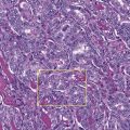
Given the diagnostic uncertainty on the initial assessment, further characterization was warranted. CEUS was performed using Definity contrast agent (Lantheus; Billerica, MA, USA). The examination followed standardized protocols for gallbladder CEUS, including continuous imaging through arterial and venous phases [ ]. CEUS demonstrated complete absence of enhancement within the gallbladder lesion throughout all phases, definitively characterizing it as a sludge ball ( Fig. 2 ).

Related posts:
 Breast cancer with medullary features shows a fast and plateau enhancement pattern on magnetic resonance images: A case report
 A rare and life-threatening case of spontaneous hemopneumothorax presenting with severe hemodynamic instability
 Postextubation negative-pressure pulmonary edema after an appendectomy
 Primary sternal osteomyelitis in an immunocompetent adolescent
 Avascular necrosis of lateral femoral condyle in a middle-aged woman from central India
 Facial nerve palsy from temporal bone metastasis in papillary carcinoma of thyroid: A case report
Stay updated, free articles. Join our Telegram channel

Full access? Get Clinical Tree


