Abstract
Mediastinal liposarcoma are rare occurrences of liposarcomas, arising from mesenchymal fat cells. They have several histological subtypes including well-differentiated, dedifferentiated, myxoid and pleomorphic. They often present with nonspecific symptoms of mass effect on nearby structures. Surgical resection is the mainstay of treatment, but high recurrence rate remains a challenge. We present the case of a 70-year-old male with a large dedifferentiated anterior mediastinal liposarcoma presenting with compressive symptoms. Coordinated efforts from radiology, histopathology and surgical teams are required for timely and accurate diagnosis and an optimized course of treatment.
Introduction
Liposarcomas are common malignant neoplasms arising in mesenchymal cells. However, mediastinal occurrence is very rare, accounting for less than 1% of all liposarcoma presentations [ ]. Patients are often diagnosed incidentally or may initially present with nonspecific symptoms arising due to mass effect and compression of nearby mediastinal structures. Liposarcomas are broadly classified by the World Health Organization (WHO) into 4 histological subtypes, including well-differentiated, dedifferentiated, myxoid/round cell and pleomorphic liposarcoma (PLS) [ ]. Due to rapid growth and a high likelihood of local recurrence of mediastinal liposarcomas, complete excision is the primary method of treatment, sometimes requiring adjuvant chemotherapy and radiotherapy supplementation depending on aggressiveness and local tissue involvement. Here, we present the case of a 70-year-old male diagnosed with dedifferentiated liposarcoma of the anterior mediastinum after presenting for exertional dyspnea and weight loss. In this article, we focus on the use of radiological imaging modalities, such as x-ray (CXR) and computed tomography (CT), coordinated with pathological findings for advanced surgical planning and optimized diagnosis.
Case presentation
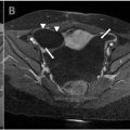
A 70-year-old male with past medical history of prostate adenocarcinoma status post prostatectomy and hyperlipidemia presented to an outside institution with symptoms of exertional dyspnea, nausea, and a 15-pound weight loss. Further review of symptoms included pain in the left shoulder blade and swelling sensation in the left neck that occurs when turning his head to the left and swallowing. Initial labs revealed elevated creatinine and hypercalcemia, and calcitonin was immediately administered. A subsequent CXR imaging of the chest was performed, and a very large anterior mediastinal mass was discovered. The initial differentials included lymphoma or thymic malignancy, and a chest computed tomography (CT) scan with contrast ( Fig. 1 ) was recommended for further evaluation. The CT findings noted multiple bulky, heterogeneously enhancing and centrally necrotic soft tissue masses in the anterior mediastinum. It measured 9.4 × 8.5 cm in the superior mediastinum and 8.6 × 0.5 cm in the left anterior mediastinum. There was subtle fat in the anterior aspect of the mass defined. The masses partially encased the proximal aspect of the left common carotid artery and subclavian artery, and abutted the brachiocephalic artery and the aortic arch. However, it did not invade the great vessels. There was rightward tracheal and superior vena cava displacement secondary to mass effect.

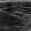
The mass was biopsied by ultrasound (US) guided needle biopsy, but the results were inconclusive, requiring surgical biopsy to help guide further management. Based on imaging, the differential diagnosis included thymic neoplasm, germ cell tumor, lymphoma, and slight concern of teratoma considering the patient’s age.
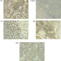
Shortly after, the patient underwent a video-assisted thoracoscopic surgical (VATS) biopsy of the mediastinal mass. Histologic sections showed a high grade pleomorphic and spindle cell sarcoma growing with a variably hyalinized and inflammatory-rich background stroma. According to the pathology report, MDM2 and CDK4 gene amplification was positive by FISH (Fluorescence in situ hybridization). Expert review of the pathology concluded that dedifferentiated liposarcoma was the best diagnosis for this soft tissue mass. Following this conclusion, the patient was referred for surgical resection of the mass.
At this point, it was suspected that the hypercalcemia was the result of the underlying malignancy, and the patient was started on Denosumab and ordered to continue on calcitonin. In addition, nephrology was consulted to address elevated creatinine levels. Subsequent imaging indicated that the mediastinal mass remains unchanged, but a small left pleural effusion and elevated left hemidiaphragm have developed. This may be due to encasement of the phrenic nerve by the large mass. Currently, he is under medical management for electrolyte derangement and nausea. The multidisciplinary surgical team is still discussing future surgical options for the patient.
Discussion
We present the case of a 70-year-old male diagnosed with a rare dedifferentiated liposarcoma of the mediastinum. Mediastinal liposarcoma is an exceedingly uncommon presentation of liposarcoma and infrequently discussed in the literature [ , ]. Liposarcomas are rare mesenchymal neoplasms that arise from soft tissue lipocytes, accounting for roughly 20% of all soft tissue sarcomas [ ]. They typically occur in the thigh or retroperitoneum in adults [ ]. Their exact etiology is unknown, and specific risk factors remain unclear, however, it is thought that radiation, specific family cancer syndromes, such as Li-Fraumeni syndrome and Werner syndrome, lymphatic trauma, and exposure to toxic chemicals may play a role in its development [ ]. Liposarcomas often remain asymptomatic until large enough to manifest compressive symptoms due to their rapid growth and displacement or infiltration of adjacent structures. Mediastinal liposarcoma may present with nonspecific symptoms of dyspnea, cough, and chest tightness [ , ].
Liposarcomas have various histological subtypes and the possibility of mixed subtypes contribute to the complexity of diagnosing specific liposarcomas. Studies have reported cases of all 4 subtypes in the mediastinum, along with few unusual variants including PLS, myxoid well-differentiated liposarcoma, thymoliposarcoma, and sclerosing high-grade liposarcoma [ , , , , ]. Dedifferentiated liposarcoma (DDLS) are most commonly location is the retroperitoneum and the extremities. DDLS often presents with compressive symptoms due to its large size at initial diagnosis. Our patient had a large anterior mediastinal mass at presentation, abutting surrounding vascular structures, making the surgical decision difficult [ , , ]. They are highly aggressive with a local recurrence rate of 40% [ ]. The aggressive nature and complexity surrounding the mixed histological subtypes of liposarcoma requires timely identification and complementary clinical, radiological, pathological, and surgical efforts to ensure the best possible outcome.
Radiological diagnostic modalities used to identify mediastinal liposarcomas, such as computed tomography (CT) and magnetic resonance imaging (MRI), are instrumental for initial diagnoses [ ]. While these studies are not specific enough to make a final diagnosis, they help narrow the differential diagnosis by evaluating the fat content of the neoplasm – higher lipomatous content is associated with lipomas, while a lower fat content is typically observed in liposarcomas. The amount of fatty tumor component will vary as in this case where the lipomatous component is small. On CT, all liposarcoma subtypes appear heterogeneous, while on MRI, high-grade well-differentiated liposarcomas show low intensity in T1 images and appear larger with thicker septa [ ]. Regardless of which imaging modality is utilized, pathology is generally required for further differentiating between the histological subtypes.
Correlation between histopathology, immunohistochemistry, and molecular testing is vital for improved differentiation between the various subtypes of liposarcomas. In addition, gene amplification has recently become a more reliable technique for classifying suspected neoplasms [ , ]. Overall, the complexity of various subtypes and insufficient knowledge of genomic structure alterations have led to significantly less confidence when pathologically diagnosing liposarcoma subtypes. So far, evaluation of the 12q13q15 gene amplifications of MDM2, CDk4, and HMGA2 allows for the distinguishment of well-differentiated/dedifferentiated liposarcomas from benign adipocytic tumors [ , ]. Distinguishing between well-differentiated and dedifferentiated is much more challenging as 10% of dedifferentiated liposarcomas are recurrences of well-differentiated liposarcomas and little is known about the mechanism or cause of the dedifferentiation [ ].
Recurrence rates for liposarcomas are very high despite attempts to treat with adjuvant chemotherapy and radiotherapy, rendering surgical resection the most effective treatment strategy for mediastinal liposarcomas [ , ]. Surgical excision of mediastinal liposarcomas as in this case can be challenging and requires careful surgical consideration and close coordination with preoperative radiological imaging for procedural safety and efficiency. Prognosis largely depends on the liposarcoma subtype and the surgical resection margin. Well-differentiated neoplasms have an excellent 5-year survival rate of about 75%-100%, with a 50% recurrence rate and relatively no risk of distal metastasis. Conversely, the more aggressive types, like myxoid or pleomorphic liposarcomas, have low survival rates of 4 – 107 months and an 80% chance of recurrence [ ]. DDLS rarely presents with distant metastasis and overall mortality has been reports between 28% and 30% at 5 years [ ]. However, as long as recurrent tumors remain excisable, multiple surgical interventions have shown the ability to locally contain liposarcomas that have not metastasized.
Conclusion
This case highlights the rare occurrence of mediastinal dedifferentiated liposarcoma—an aggressive and challenging malignancy that presents unique diagnostic and treatment challenges. Despite being an uncommon presentation of liposarcoma, the initial diagnosis was supported by timely and correlated radiological and pathological findings. Though typically associated with a high risk of recurrence, surgical resection remains the mainstay of treatment. This case emphasizes the importance of a multidisciplinary approach involving radiology, pathology, and surgical coordination to ensure the most effective treatment strategy. Continued research needs to be completed to further our understanding of the dedifferentiation mechanism and genomic amplification for improved diagnostic confidence.
Patient consent
The patient reported in the manuscript signed the informed consent/authorization for participation in research, which includes the permission to use data collected in future research projects such as the presented case details and images used in this manuscript.
Competing Interests: The authors declare that they have no known competing financial interests or personal relationships that could have appeared to influence the work reported in this paper.
Acknowledgments: There was no funding associated with this report.
References
Related posts:
 Breast cancer with medullary features shows a fast and plateau enhancement pattern on magnetic resonance images: A case report
 Ovarian metastasis from lobular breast carcinoma: A case report with review of literature
 Invasive lobular carcinoma with metastasis to the pectoralis muscle
 A rare and life-threatening case of spontaneous hemopneumothorax presenting with severe hemodynamic instability
 Postextubation negative-pressure pulmonary edema after an appendectomy
 Uncovering a peritoneal inclusion cyst after surgery: A case report
Stay updated, free articles. Join our Telegram channel

Full access? Get Clinical Tree


