Abstract
Demons-Meigs (DM) syndrome is characterized by the association of a benign ovarian tumor most often a fibroma or fibrothecoma with pleural and intraperitoneal effusions. It is a rare pathological entity. We report the case of a 40-year-old female patient with typical DM syndrome, characterized by the coexistence of pleural and intraperitoneal effusions, an ovarian mass, and a CA-125 level of 633 IU/ml. Laparotomy revealed abundant ascites and a large right ovarian mass, which was subsequently removed. Histopathological examination confirmed an ovarian fibroma. DM syndrome has a favorable prognosis, and treatment is primarily based on the removal of the ovarian tumor, without the need for chemotherapy or other therapeutic approaches.
Introduction
DM syndrome is a condition involving a benign pelvic tumor, typically an ovarian fibroma, associated with pleural and/or peritoneal effusions [ ]. It remains an exceptionally rare condition, and its pathophysiology is poorly understood [ , , ]. Imaging techniques such as ultrasound, CT, and MRI play a crucial role in diagnosing DM syndrome by identifying the benign ovarian mass and associated effusions [ , ]. Fibromas and fibrothecomas are the most commonly reported tumors associated with this syndrome. However, before histological examination of the tumor, this rare syndrome often raises concerns about malignancy due to the tumor’s size, the presence of ascites, and significantly elevated CA-125 levels [ , , ].
Case presentation
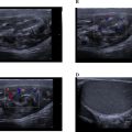
We report the case of a 40-year-old female patient with no significant medical history who presented with abdominal distension, pelvic heaviness, right-sided chest pain, and dyspnea. Clinical examination revealed bilateral pleural effusion, abdominal distension, and a positive fluid wave test. Ultrasound identified a solid-cystic pelvic mass measuring 26 cm in diameter, accompanied by abundant ascites. A thoracic-abdominopelvic CT scan showed a large right ovarian mass with lobulated contours, approximately 28 cm in diameter. The mass exhibited minimal enhancement after contrast administration, consistent with its fibrous nature, and contained calcifications and hypodense areas. No invasion of adjacent structures was observed, and the scan confirmed abundant bilateral pleural and intraperitoneal effusions with no distant metastases ( Fig. 1 ).

MRI revealed a large, well-circumscribed, oval-shaped right latero-uterine mass adjacent to the ovary, hypointense on T2-weighted imaging, nonrestrictive on diffusion, and weakly enhanced after contrast administration ( Fig. 2 ). The patient underwent laparotomy, which revealed a large right ovarian mass ( Fig. 3 ) and a significant volume of transudative ascites. The tumor was removed via oophorectomy and omentectomy, and 17 liters of ascites were evacuated. Histological and immunohistochemical findings confirmed an ovarian fibroma ( Fig. 4 ). The postoperative course was uneventful, with complete resolution of pleural and peritoneal effusions observed immediately after surgery, without the need for chemotherapy. CA-125 levels were significantly reduced 6 months after the procedure.

Related posts:
 Breast cancer with medullary features shows a fast and plateau enhancement pattern on magnetic resonance images: A case report
 A rare and life-threatening case of spontaneous hemopneumothorax presenting with severe hemodynamic instability
 Avascular necrosis of lateral femoral condyle in a middle-aged woman from central India
 A very rare case of ileocolic and appendiceal intussusception with acute appendicitis
 Rare case of left epididymo-orchitis complicated by pampiniform plexus thrombosis: A case report
 Hepatic and extra-hepatic hydatid cysts: A case series of radiological and clinical insights
Stay updated, free articles. Join our Telegram channel

Full access? Get Clinical Tree


