KEY FACTS
Terminology
- •
Well-defined histology: Papillary and follicular carcinoma
Imaging
- •
Papillary carcinoma (~ 80% of thyroid cancer)
- ○
Majority are ill-defined with irregular outlines
- ○
10-20% multifocal, 70% solid; 77-90% hypoechoic
- ○
Calcifications: Punctate small echogenic foci highly specific; larger calcifications also concerning
- ○
Cystic variant: Rare, eccentric polypoid solid vascular nodule within cyst ± microcalcifications
- ○
Nodes predominantly hyperechoic (80%) compared to muscles; 50% with punctate microcalcification; cystic change in 25%
- ○
- •
Follicular carcinoma (~ 10% of thyroid cancer)
- ○
Ill-defined solid tumor; hypoechoic, heterogeneous
- ○
- •
Large tumors invade strap muscles, esophagus, trachea, recurrent laryngeal nerve, neck vessels
- •
Color Doppler: Profuse chaotic vascularity within nodule or in wall, and septa of partially cystic tumors
Top Differential Diagnoses
- •
Multinodular goiter
- •
Thyroid adenoma
- •
Aggressive thyroid carcinomas and metastases
Clinical Issues
- •
Painless, palpable thyroid nodule or incidental finding on imaging
- •
Rapid growth of thyroid mass, extrathyroidal hard nodes, hoarseness; history of radiation exposure
- •
Low mortality from malignancy; 20-year survival rate: 90% papillary, 75% follicular
- •
Peak incidence in 3rd and 4th decade; F:M = 3:1
Scanning Tips
- •
Look for extrathyroidal extension by evaluating capsule and mobility during swallowing; evaluate nodal chains
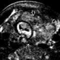
 is taller than it is wide, with lobulated margins, and hypoechoic relative to normal thyroid
  .
.
 shows color flow in the center of the tumor.
 is markedly hypoechoic relative to normal thyroid
  .
.
 is poorly marginated and is hypoechoic in comparison to the thyroid with multiple echogenic foci.
 with a thin calcified incomplete rim. There are numerous small punctate echogenic foci
  .
.


