Duodenal Carcinoma
Michael P. Federle, MD, FACR
Key Facts
Imaging
Irregular intraluminal mass or “apple core” lesion at or distal to ampulla of Vater
Irregular thickening of duodenal wall
Concentric narrowing of duodenal lumen
Polypoid intraluminal mass
Local lymphadenopathy and local infiltration
Biliary ± pancreatic duct dilatation
With periampullary tumors
Liver ± peritoneal metastases
Top Differential Diagnoses
Pancreatic ductal carcinoma
Ampullary carcinoma
Intestinal metastases and lymphoma
Malignant GI stromal tumor
Duodenal ulcer
Crohn disease
Tuberculosis
Annular pancreas
Clinical Issues
Other signs/symptoms
Nausea and vomiting, weight loss, anemia, upper GI bleed
Periampullary tumors may present with jaundice
Rare: Represents < 1% of all gastrointestinal neoplasms
Diagnostic Checklist
Most duodenal carcinomas cause focal stenoses or obstruction; large mass with cavitation is often lymphoma or GIST
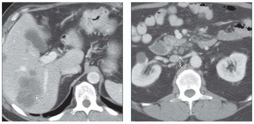 (Left) Axial CECT in a 60-year-old man with weight loss and early satiety shows obvious liver metastases  . (Right) Axial CECT in the same patient also shows paraduodenal lymph node metastases . (Right) Axial CECT in the same patient also shows paraduodenal lymph node metastases  . . |
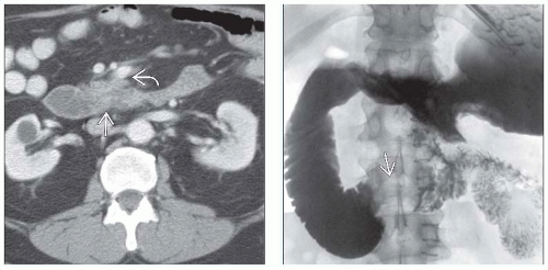 (Left) Axial CECT in the same patient shows the relatively subtle mass that narrows the 3rd portion of the duodenum  . There is also a subtle extension of tumor along the superior mesenteric vessels . There is also a subtle extension of tumor along the superior mesenteric vessels  . (Right) Film from an upper GI series in the same patient shows the duodenal carcinoma more clearly . (Right) Film from an upper GI series in the same patient shows the duodenal carcinoma more clearly  . Note the “shoulder” or abrupt transition to tumor at its proximal extent. The lumen of the more proximal duodenum is dilated. . Note the “shoulder” or abrupt transition to tumor at its proximal extent. The lumen of the more proximal duodenum is dilated. |
TERMINOLOGY
Abbreviations
Duodenal carcinoma (CA), duodenal adenocarcinoma
Definitions
Primary malignant neoplasm arising in duodenal mucosa
IMAGING
General Features
Best diagnostic clue
Irregular intraluminal mass or “apple core” lesion at or distal to ampulla of Vater
Location
15% in 1st portion of duodenum
40% in 2nd portion of duodenum
45% in distal duodenum
Size
Usually < 8 cm
Morphology
Polypoid, ulcerated, or annular constricting mass
Intraluminal mass with numerous frond-like projections for carcinomas arising in villous tumors
Fluoroscopic Findings
May have various appearances
Ulcerated mass
Polypoid mass
Annular constricting “apple core” lesion
“Soap bubble” reticulated pattern for villous tumors
CT Findings
CECT
Discrete mass or irregular thickening of duodenal wall
Concentric narrowing of duodenal lumen
Polypoid intraluminal mass
Local lymphadenopathy
Infiltration of adjacent fat
Biliary ± pancreatic duct dilatation
With periampullary tumors
Liver ± peritoneal metastases
MR Findings
MRCP
May see pancreatic or biliary ductal dilatation with periampullary duodenal carcinomas
Ultrasonographic Findings
Grayscale ultrasoundRelated posts:

Stay updated, free articles. Join our Telegram channel

Full access? Get Clinical Tree


