KEY FACTS
Imaging
- •
Multiple causes; can be isolated or syndromic
- •
Autosomal recessive polycystic kidney disease is single gene disorder
- ○
Large, diffusely hyperechoic kidneys most common
- ○
May see discrete cysts or areas of tubular ectasia
- ○
Sometimes just echogenic medullary pyramids
- ○
- •
Obstructive cystic dysplasia
- ○
Microscopic cysts develop due to chronic obstruction
- ○
Multiple interfaces → increased echogenicity
- ○
Macroscopic cortical cysts usually present
- ○
- •
Trisomy 13
- ○
Echogenic kidneys seen in 50%
- ○
Look for holoprosencephaly, cyclopia, proboscis, midline facial cleft, polydactyly, congenital heart disease
- ○
Early-onset fetal growth restriction
- ○
- •
Meckel-Gruber syndrome
- ○
Triad of findings: 2 needed to make diagnosis
- –
Renal cystic dysplasia in > 90%
- –
Encephalocele in 60-80%
- –
Polydactyly in 55-75%
- –
- ○
Scanning Tips
- •
Echogenic kidneys are brighter than liver
- •
Use high-resolution transducer to look for subtle cystic change/tubular ectasia
- •
Use vaginal transducer for better resolution in 1st trimester or with breech presentation
- •
If fluid is low, verify that membrane rupture has been ruled out: Relative contraindication to transvaginal ultrasound (TVUS) due to risk of introducing infection
- •
Look for other anomalies, more likely syndromal diagnosis
- •
Oligohydramnios may cause pulmonary hypoplasia
- ○
Measure chest:heart ratios to document small chest size
- ○
Normally, heart circumference is 1/2 of chest; heart area is 1/3 of chest
- ○
 . The left measured 3.7 cm (> 90th percentile for gestational age). In this case, amniotic fluid volume was normal throughout pregnancy, and the diagnosis of autosomal recessive polycystic kidney disease (ARPKD) was established at birth.
 in large kidneys. Oligohydramnios was present in this case as well.
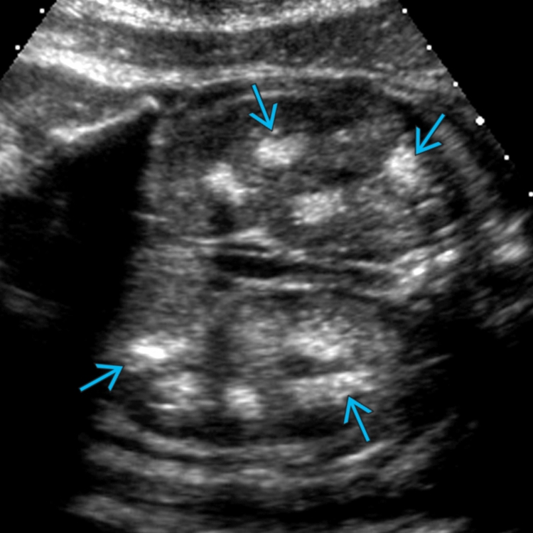
 in the left upper pole of a fetus with ARPKD. The kidneys are very large (calipers measure a 9-cm left kidney), and the medullary pyramids
  are echogenic.
 and focal, upper pole tubular ectasia
  .
.
 and a left, liver-up diaphragmatic hernia
  . Amniocentesis revealed trisomy 13.


