Ectopic Pregnancy
Niyati Mukherjee
CLINICAL HISTORY
36-year-old female presenting to the emergency department with diffuse abdominal pain. Last menstrual period (LMP) was 9 weeks ago. She recently underwent dilation and curettage (D&C) for suspected ectopic pregnancy with no intrauterine chorionic villi detected during the procedure, and is currently on methotrexate. Transabdominal ultrasound was performed initially; the exam was terminated prior to an attempt at transvaginal ultrasound based on the preliminary findings.
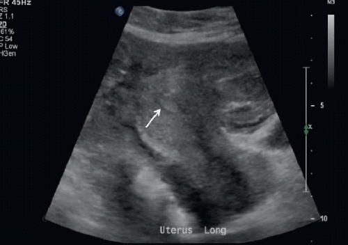 FIGURE 15A |
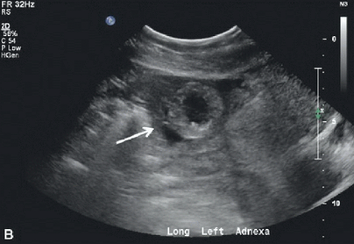 FIGURE 15B |
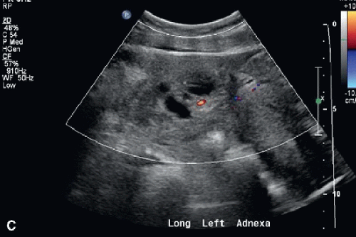 FIGURE 15C |
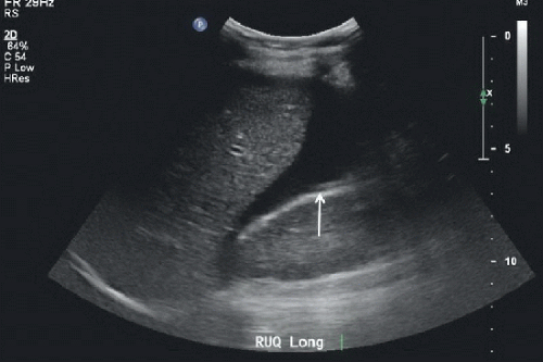 FIGURE 15D |
FINDINGS
Figure 15A: Transabdominal US longitudinal image of the uterus. The endometrial stripe is visualized (arrow). No intrauterine pregnancy is identified. Figures 15B and 15C: Transabdominal US longitudinal images of the left adnexa, with and without color Doppler. An echogenic ring with surrounding fluid is seen in the left adnexa (arrow). This is the “tubal ring sign,” worrisome for tubal ectopic pregnancy. Mild vascularity is noted. Normal ovaries are not identified on imaging of the adnexae. Figure 15D: Transabdominal US longitudinal image of the right upper quadrant. Free fluid is seen in the upper abdomen (arrow), as well as the pelvis as seen in Figure 15B, which is worrisome for a ruptured ectopic pregnancy.
DIFFERENTIAL DIAGNOSIS
Tubal ectopic pregnancy with rupture.
Exophytic corpus luteum cyst with associated hemorrhage or rupture.
DIAGNOSIS
Related posts:


Stay updated, free articles. Join our Telegram channel

Full access? Get Clinical Tree


