Abstract
Background
Traditionally, transforaminal steroid injection is performed in the management of cervical radiculopathy in medical failure treatment but carried a true risk of catastrophic complication. Another approach currently used is to perform intra-articular facet steroid injection to reach the epidural space.
Purpose
The aim of this study was to describe the evolution of symptoms following intra-articular facet steroid injection in cervical radiculopathy.
Material and methods
We conducted a retrospective study. We assessed all patients who had a CT-guided intra-articular facet steroid injection in our center (xx, xx, xx) from December 2015 to February 2021. Cervical MR pretherapeutic images were analyzed and classified according to cervical pain etiology: uncodiscarthrosis, disk herniation or congestive cervical posterior osteo-arthritis. All patients had clinical initial evaluation and then follow-up at 1 and 6 months. Pain severity was rated on a visual analog scale and expressed as a percentage of improvement.
Results
Ninety-three patients were included. There were 56 patients with uncodiscarthrosis, 29 with a disk herniation and 8 with a cervical posterior congestive osteoarthritis. A significant improvement of the visual analog scale percentage was found for all patient at 1 and 6 months ( p < 0.01). Visual analog scale percentage improvement was about 50 % for all etiologies. For all patients, no severe complications were reported.
Conclusion
Intra-articular facet steroid injection may be considered for the treatment of cervical radiculopathy when other medical treatments have failed.
1
Introduction
Cervical radiculopathy is a frequent and common pathology that involves pain perceived in the neck with radiation to the upper extremity [ ]. This clinical condition results from the compression of a cervical root or an inflammation process surrounding the root. In most cases, it affects lower cervical spine (from C4 to C7) [ ]. Common causes are differentiated from symptomatic causes (hematoma, infection and inflammatory pathology). The two main etiologies of common cervical radiculopathy are uncodiscarthrosis and disc herniation [ ]. Uncodiscarthrosis is the most frequent (70 %) [ ]. Another under-studied cause is congestive posterior articular osteoarthritis. Indeed, 80 % of congestive flare-up of posterior articular osteoarthritis induce an ipsilateral cervical radiculopathy. Prevalence of congestive posterior osteoarthritis in CR is about 10 % [ ].
Non-invasive management includes relative rest, oral analgesics, non-steroidal anti-inflammatory drugs and physical therapy with inconclusive evidence for their effectiveness [ , ]. For patients with cervical radiculopathy, improvements in pain and function tend to occur only within several months, with significant disability and economic impact [ ]. In case of failure of these treatments, image-guided nerve root infiltration of steroids can be performed. However, surgery does not have any place in first line; indeed, the benefit / risk balance is low with a high risk of complication [ ]. Traditionally, transforaminal steroid injection (TFSI) or even interlaminar epidural injection were used. Theoretically, TFSI is supposed to deliver a selective and higher concentration of steroids near the cervical nerve root and in the ventral epidural space [ , ]. Nevertheless, TFSI seems to be responsible for extremely rare but catastrophic complications. Reports of quadriparesis, stroke and even death following TFSI began appearing in the early 2000s [ ]. Intra-articular facet steroid injection (IFSI) is an alternative technique available to deliver steroids close to the cervical nerve root. Indeed, the cervical posterior facet joint is a way of diffusion to the foraminal space, through the retrodural space also called space of Okada [ ]. This method, little studied in the literature, seems interesting because it is technically easier and presents fewer neurological risks. A recent study showed an equivalent efficacity of IFSI and TFSI in this indication, but there were some limits, in particular, the clinical outcomes were only evaluated early at one month [ ]. Another study demonstrated the efficacity of IFSI but the infiltrations were performed with fluoroscopy [ ], which induces a risk of navigation error. Moreover, in these two studies the cause of the cervical radiculopathywas not investigated. Therefore, this study aimed at evaluating the efficacity of IFSI with CT guidance in patients with cervical radiculopathy. A secondary objective was to better identify the effectiveness of ISFI according to the etiology.
2
Material and methods
2.1
Participants
We conducted a single-centered retrospective study. We identified all patients who underwent an IFSI from December 2015 to February 2021 in our center (xx, xx, xx) using our local pictorial archive and communication system (Carestream 3.2, Carestream Health, Rochester, New York). All patients gave oral consent before IFSI. To ensure full consistency of the level injected and the most plausible nociceptive lesion, all patients had a consultation in a multidisciplinary team meeting, during which the clinical presentation and imaging were reviewed by a multidisciplinary team of spine specialists (i.e. radiologists, rheumatologists, spine surgeons, physiatrists) with at least 5-year experience in the management of spine disorders. If several anatomical lesions were present, the patient was classified in the group of the lesion most consistent with the clinical presentation and that was considered for injection. For the purpose of our study, patients with mixed lesions (i.e., combination of uncarthrosis, disk herniation and/or posterior impingement due to a congestive posterior articular) at the same level were not included. The eligibility criteria were patients over 18 years old with a common cervical radiculopathy who underwent IFSI under CT guidance in our center. Only patients with a single site of injection were included. We excluded patients who did not perform cervical MR in the preceding six months, or who did not get complete follow-up (at least 6 months).
2.2
MR analysis
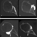
Cervical MR images were analyzed on our institutional imaging server. Examinations were not all performed in our center, so MR imaging protocol was not similar for all patients. A single junior radiologist with one year of experience in musculoskeletal imaging reviewed all exams. MR imaging all contained at least a sagittal T2-weighted sequence, a sagittal T1-weighted and an axial T2-weighted acquisition. Most examinations specifically encompassed an inflammatory sequence (T2-weighted sequence with fat suppression technique), such as a sagittal T2-Dixon, sagittal STIR or coronal STIR. MR imaging was reviewed to confirm the presence of uncodiscarthrosis, disk herniation or congestive posterior articular osteoarthritis at the level of the involved cervical nerve. Three different groups were defined according to the presence of anatomical changes on MRI including at least a sagittal T2-, a sagittal T1- and an axial T2-weighted sequence, consistent with clinical presentation, as assessed during a multidisciplinary team meeting. Group A ( Fig. 1 ) was defined by the presence of osteoarthritic changes involving the uncus, group B ( Fig. 2 ) by the presence of a herniated disc and group C ( Fig. 3 ) by the presence of congestive osteoarthritic changes involving the facet joint. When an inflammatory sequence was missing, congestive posterior osteoarthritis changes were defined as facet joint osteoarthritic changes associated with joint hypointense signal in sagittal T1-weighted sequence and hyperintense signal in sagittal T2-weighted sequence.


2.3
Intervention
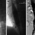
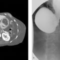
The intervention was realized by three radiologists from our center, all with experience in musculoskeletal imaging ranging from 3 to 15 years. We performed a checklist safety before all interventions: iodine-based contrast allergy, current infection, coagulopathy and absence of cervical surgery. All patients were installed in the prone position. The injection was performed in sterile conditions by a standardized technique [ ] under CT guidance (Somaton Definition Flash, Siemens, Germany). The appropriate entry site was marked on the skin after spotting. Then a 22-ga 3,5-inch spinal needle was advanced by using intermittent CT-controlled. Once the needle was in the appropriate location, 0.5–1 ml of iodinated contrast in the cervical facet joint was injected. The iodinated contrast injected was Visipaque 320 mg iodixanol/ml. The foraminal space could be opacified in some cases after contrast injection in the cervical facet joint ( Fig. 4 ). A final CT-control was then performed to exclude an intra-vascular position and to confirm the adequate intra-articular distribution of the contrast. Then, 1 ml of dexamethasone sodium phosphate 10 mg/ml was injected, and the needle was withdrawn. The infiltration was performed on the ipsilateral posterior cervical facet of the cervical radiculopathy. No local anesthetics were used during the procedure. The patient was systematically observed for 30 min after the intervention.

2.4
Outcomes
The indication of injection and the follow-up were performed by physicians from the physical medicine and rehabilitation department of our center. The clinical outcome measure was pain severity rated on a visual analog scale (VAS): defined by a line extending from 0 (no pain) to 10 (worst pain imaginable). All patients were evaluated with this VAS three times by physicians: before the intervention, then 0-1 month and 1–6 months after the intervention. Pain assessments before and after could be performed either by the treating physician and/or by the radiologist, in the context of health care. The improvement of the pain compared to the initial consultation was expressed using a percentage improvement of the VAS (VAS%). VAS% was calculated in the first month after the infiltration (compared with the initial consultation) and between 1 and 6 months after the infiltration (compared with the initial consultation too). For all patients, we also specified if they took analgesic treatment including morphine and if they did or did not have a professional activity during their pathology (classical factors predictors of poor response). Patients in retirement were considered in the professional activity group. Complications were also noted: hematoma, infection and neurological accident. Clinical outcomes were analyzed in blind of the MRI results.
2.5
Statistical analysis
The data analyses were performed with RStudio software (RStudio, Inc, version 1.2.5033). Quantitative continuous variables results were presented as means with standard deviation or median with range according to the distribution and qualitative variable by frequency (%). The normal distribution of variables was tested using a Shapiro test. Quantitative variables were tested using a Wilcoxon test or a student t -test according to their distribution. For analyses with more than two groups, a one-way ANOVA analysis was used with Tukey’s multiple comparisons. For all analyses, significant results were obtained by p < 0.05.
2.6
Ethical consideration
Because our research was retrospective, based on data already collected in the context of health care, which therefore, by definition, did not involve human beings directly, it did not fall under the Jardé Law of March 5th, 2012 and its application decree (No. 2016-1537) related to research involving the human person in France (https://www.legifrance.gouv.fr/jorf/id/JORFTEXT000025441587, accessed on February 8th, 2023). Therefore, a formal approval from an Institutional Review Board (IRB) is not mandatory. Further, in our experience, requesting an authorization of an ethical committee (e.g., Cerim), after the research was conducted, is usually considered as inappropriate by the committees. For this reason, we did not request this authorization for the purpose of the present manuscript. Finally, all patients, in the context of health care offered in our institution, were fully informed that data could be retrieved from their medical records for research purpose and were free to decline at any time.
3
Results
3.1
Participants
Overall, 401 patients with intra-articular cervical facet joint steroid infiltrations were initially eligible; 308 patients were excluded: 110 did not have a complete clinical follow-up and 198 did not have MR imaging available or achieve within the 6 months. Finally, 93 patients were included in this study ( Table 1 ). It was the first IFSI for all participants.
Related posts:
 Glenoid morphology variation between patients with hypermobile shoulder joints and controls: Identification of hyperlaxity-related morphologic bone changes
 Uterine Fibroid Embolization
 Anesthetic Care for Interventional Neuroradiological Procedures
 Migrated Superior Vena Cava Stent Repositioned Using a Balloon and Loop Snare Combination
 Part 1 Preprocedural Cases
 25 Islet Cell Transplantation
Stay updated, free articles. Join our Telegram channel

Full access? Get Clinical Tree


