Abstract
Objective
Measurement of thenar muscle elasticity by ultrasound shear wave elastography (SWE) may be useful for the diagnosis and evaluation of carpal tunnel syndrome (CTS), but there is a paucity of information on SWE of the thenar muscles in patients with CTS. The purpose of this study was to investigate the elasticity of the thenar muscles in patients with CTS.
Methods
Twenty-two adult patients with a referral diagnosis of CTS (27 hands) and 20 healthy volunteers as a control (20 dominant hands) participated in this study. The elastic modulus of the thenar muscles was measured with SWE in two conditions, rest and pinch. The elastic modulus and percent change between the two conditions for each muscle were compared between groups.
Results
The elastic modulus of the abductor pollicis brevis (APB) in the patient group was lower than that in the control group at rest (20.6 ± 5.6 kPa vs. 25.0 ± 8.5 kPa; p = 0.034). However, the elastic modulus (87.9 ± 49.0 kPa vs. 62.5 ± 28.5 kPa; p = 0.044) and percent change (373.3 ± 336.4% vs. 169.2 ± 116.0%; p = 0.006) of the APB in the patient group were higher than those in the control group with pinch. For the opponens pollicis and adductor pollicis, there was no significant difference in the elastic modulus between groups.
Conclusion
Patients with CTS showed differences in SWE of the APB. Future studies need to investigate the accuracy of SWE in diagnosing CTS and assessing prognosis.
Introduction
Carpal tunnel syndrome (CTS) is one of the most prevalent conditions in the hand [ , ]. It is commonly described as a compression neuropathy, affecting the median nerve at the level of the transverse carpal ligament. Electrodiagnostic studies (EDX) are commonly used to evaluate and diagnose patients with suspected CTS [ , ]. EDX can be perceived as painful and a cause of anxiety, sometimes resulting in incomplete or canceled testing [ , ].
Neuromuscular ultrasound (NMUS) is becoming more widely used for the non-invasive evaluation and diagnosis of CTS as it provides anatomic information complementary to the physiological information provided by EDX. Expert consensus strongly recommends its application in a number of clinical scenarios related to CTS [ ]. A recent study demonstrated that patients with CTS prefer NMUS over EDX for the diagnosis of CTS [ ]. To date, however, there have not been NMUS parameters that strongly correlate with patient-reported symptom severity [ ] or severity as defined by EDX criteria [ ], especially in the elderly population [ ]. Prior NMUS studies have focused on size changes in the thenar muscles, such as thickness and cross-sectional area [ ]. Several studies have also evaluated muscle echo intensity to detect denervation-related changes in the thenar muscles [ , ]. While these conventional NMUS technologies can provide information on morphology (e.g., size) and compositional features (e.g., fibrosis and fatty infiltration) due to denervation, they do not directly assess the mechanical properties of the thenar muscles.
Ultrasound shear wave elastography (SWE) is an imaging modality that evaluates the elasticity of soft tissues by measuring the speed of propagation of a shear wave through these tissues. SWE may aid in early disease detection when abnormality cannot yet be visualized on conventional NMUS [ ]. Early SWE research studied nerve stiffness in compressive neuropathies [ ]. However, a recent review article concluded that elastography evaluation of the median nerve is not suitable as an independent diagnostic tool, although it may provide additional insight into the severity of CTS [ ]. A recent study using SWE on the muscle demonstrated that the elasticity of the thenar muscles of patients with CTS was higher than that of healthy controls, and that there was a negative correlation between elasticity and severity of CTS [ ]. However, this study measured elasticity of the thenar eminence rather than individual thenar muscles and only measured elasticity at rest, thereby not taking full advantage of the capabilities of SWE. In order to understand whether SWE can be used in the future for diagnosis and prognosis assessment of CTS, it is necessary to characterize the elasticity of the thenar muscle of patients with CTS in more detail. The purpose of this study was to investigate the characteristics of elasticity of individual thenar muscles in patients with CTS by comparing the elastic modulus between patients with CTS and healthy volunteers both at rest and during pinch. We hypothesized that the elastic modulus in the median nerve–innervated thenar muscles would differ in patients with CTS from healthy volunteers under both measurement conditions.
Materials and methods
Participants
Twenty-two adult patients with a referral diagnosis of CTS (27 hands) were included in this prospective case-control study. Patients were selected from among those referred to our EDX laboratory for diagnostic evaluation for CTS. The criteria used to diagnose CTS was a combination of history, symptoms and EDX. In addition, 20 healthy, asymptomatic volunteers (20 dominant hands) were recruited from staff working in our institution as a control group. The following disorders were criteria for exclusion: history of cervical radiculopathy, rheumatoid arthritis, flexor tendinitis, gout, hemodialysis, sarcoidosis, peripheral nerve disease, amyloidosis and traumatic injuries to the ipsilateral arm. These exclusion criteria were identified based on self-report and medical record review. Participants were also excluded from this study if they had a history of surgery, fractures of the wrist or hand, or disorders to the musculoskeletal system. The study was approved by our Institutional Review Board (06-002950) and all participants provided written informed consent prior to the study.
Ultrasound measurement
A MACH 30 Ultrasound System with an SL18-5 MHz linear transducer (SuperSonic Imagine Co. Ltd, headquarters is in Aix-en-Provence, FRANCE (US office location is Seattle, WA)) was used for SWE measurement of the thenar muscles including the abductor pollicis brevis (APB), opponens pollicis (OPP) and adductor pollicis (ADP) of the affected hand or dominant hand for the patient and control group, respectively. Measurements were performed for two conditions, rest and pinch. Sufficient transmission gel was placed between the transducer and the skin to minimize muscle deformation from direct pressure from the transducer. Participants were seated with the arm positioned on an adjacent table with the forearm neutral, the elbow flexed to 60 degrees, and the wrist in the neutral position. Participants were instructed to completely relax their thumbs in a rest condition. To measure SWE with pinch, participants were instructed to hold 2 kg on a pinch meter (Digital Pressure Gauge BL-500, Baseline Corp., Golden, CO, USA) while watching the pinch meter display with a lateral pinch. The SWE measurement of the ADP was not made with pinch because the transducer could not be placed in the palm, over this muscle, during pinch. The examiner was not blinded to the ultrasound measurement.
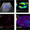
All SWE images were acquired in the long axis to the muscle fibers from the palmar surface of the hand. For the APB and OPP, the transducer was longitudinally placed on the first metacarpal (MC1) so that the proximal and distal epiphyseal region of the MC1 were visible. For the APB, the transducer was tilted to the ulnar side of the MC1 until the APB was clearly identified ( Fig. 1 a, 1 b) [ ]. For the OPP, the transducer was rotated so that the transducer matched the muscle fiber orientation of the OPP ( Fig. 1 a, 1 c). For the ADP, the transducer was placed on the palm along the long axis of the ADP at the position where the second and third metacarpals (MC2 and MC3, respectively) were visualized ( Fig. 1 a, 1 d).

Software embedded in the SWE was used for elastic modulus analysis. For the APB and OPP, three adjacent regions of interest (ROIs) with a diameter equal to the muscle diameter were placed at the site of maximum muscle thickness ( Fig. 1 a, 1 b) [ ]. For the ADP, three adjacent ROIs with a diameter equal to the muscle diameter were placed above the MC2 base ( Fig. 1 c). The SWE measurement was performed twice for each muscle. The median of the three ROIs was defined as the representative value of the image. The mean value of the two images was regarded as the elastic modulus of the tested muscle. Prior to this study, intra- and inter-rater reliability of this measurement method was tested in the APB and OPP, and intra-rater reliability was shown to be moderate and good, respectively. Inter-rater reliability was shown to be moderate for both muscles [ ]. Reliability of this measurement method has not been tested for the ADP.
CTS is often associated with concurrent thumb carpometacarpal osteoarthritis (CMC OA) [ , ], and OPP fatty infiltration (OPP FI) is a frequent finding in patients with CMC OA [ ]. Thus, after the SWE measurement, the existence of CMC OA and OPP FI were qualitatively evaluated using brightness mode in the same ultrasound system in both groups. For CMC OA, which was defined by the presence of osteophytes, an experienced physician-sonographer evaluated the CMC joint in the long axis [ ]. For OPP FI, the same physician-sonographer qualitatively evaluated the echo intensity by long- and short-axis scanning [ , ], and FI was defined when hyper echo intensity consistent with fatty infiltration was found [ , ].
In addition, the cross-sectional area (CSA) of the median nerve was also measured in brightness mode at the level of the wrist joint in the patient group during electrodiagnostic evaluation.
Electrodiagnostic studies
Electromyography and nerve conduction studies were performed in the patient group using Sierra Summit (Cadwell Industries Inc., Beloit, WI, USA) using the laboratory’s standard techniques and previously established normal values with the hand warmed to a surface temperature of ≥32 °C. In accordance with criteria of the authors’ institution, the median motor distal latency recorded at the APB ≥4.5 ms, median sensory antidromic (recorded over digit two) distal latency ≥3.6 ms, or median orthodromic (palmar) ≥2.3 ms or ≥0.4 ms difference in median compared with ulnar palmar orthodromic sensory distal latencies were considered abnormal. Electrophysiologic severity of median neuropathy, if present, was classified as mild (prolonged sensory distal latency), moderate (prolonged sensory and motor distal latencies) or severe (absent sensory response or reduced amplitude of the median motor compound muscle action potential) [ ]. Control subjects were not evaluated electrodiagnostically.
Statistical analysis
We estimated that a sample size of 20 hands in each group were required based on our preliminary measurement (0.9–1.0 effect size, 0.05 a-level and a 0.8 desired power level). All data were analyzed using SPSS Statistics Ver. 28.0 (IBM, Armonk, NY, USA). Mean age was compared between groups using an independent t -test. Sex distribution, existence of CMC OA and existence of OPP FI were compared between groups using a Χ 2 test. The elastic modulus of the APB and OPP was compared using a two-way analysis of variance (ANOVA) with group (patient vs. control) as an independent factor and condition (rest vs. pinch) as a repeated factor. When a significant interaction was observed, separate comparisons between groups were performed using two-sample t -tests. In addition, the percent change of the elastic modulus was calculated, defined in eqn (1 ) below. This is because the muscle elastic modulus at rest may differ between groups, and normalizing by the elastic modulus at rest more clearly assesses the change of the elastic modulus with pinch.
percentchange=Epinch−ErestErest×100%
Related posts:
 WFUMB Commentary Paper on Artificial intelligence in Medical Ultrasound Imaging
 Quantitative Liver Fat Assessment by Handheld Point-of-Care Ultrasound: A Technical Implementation and Pilot Study in Adults
 Automatic Segmentation of Sylvian Fissure in Brain Ultrasound Images of Pre-Term Infants Using Deep Learning Models
 A Feasible Method for Vein Puncture and Drug Administration in Rats: Ultrasound-guided Internal Jugular Vein Puncture
 Noninvasive Assessment of Liver Fibrosis in Patients With Iron Overload
 Assessment of Coronary Microcirculation with High Frame-Rate Contrast-Enhanced Echocardiography
Stay updated, free articles. Join our Telegram channel

Full access? Get Clinical Tree


