Empyema
Peter J. Noone
Katherine R. Birchard
CLINICAL HISTORY
39-year-old female with malaise.
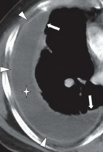 FIGURE 22A |
FINDINGS
Figure 22A: Axial contrast-enhanced CT of right hemithorax shows thickened enhancing visceral pleura (arrows) and thickened enhancing parietal pleura (arrowheads), surrounding a fluid collection (star) that is higher in attenuation than simple fluid.
DIFFERENTIAL DIAGNOSIS
Empyema, simple pleural effusion, hydropneumothorax, pulmonary abscess, chylothorax.
DIAGNOSIS
Empyema.
DISCUSSION
An empyema is a collection of purulent fluid in the pleural space.1 Empyemas are loculated, as opposed to a simple transudative effusion, which most often layers dependently in the pleural space. On CT, the purulent fluid is surrounded by thickened and enhancing pleura, known as the “split pleura sign,” also illustrated in the Figure 22A. Findings of chronic empyema include increased thickness of adjacent extrapleural fat.1 Diagnosis of empyema can be confirmed with thoracentesis and return of purulent fluid. Analysis of fluid typically reveals elevated neutrophil count, pH below 7.0, or glucose below 40 mg/dL. Streptococcus pneumoniae and Staphylococcus aureus are commonly causative agents.2
Related posts:


Stay updated, free articles. Join our Telegram channel

Full access? Get Clinical Tree


