Abstract
Port catheters are widely used today due to increase in oncology cases, while they offer ease of use, they also bring various complications. These complications can range from simple issues to life-threatening ones. Port catheter fractures are rare complications, occurring in less than 1% of patients. The most common complaint in the present study was pain at the site of the subclavian vein puncture when fluid was administered from the port hub. Simple imaging such as chest X-rays can reveal the presence of the catheter fracture and migration. Pinch-off syndrome (POS) is one of the most significant causes of this condition. It is characterized by the catheter fracture compressed between the clavicle and the first rib. The incidence of POS ranges from 0.4% to 5%, with the clinical presentation varying widely from asymptomatic cases to serious complications such as cardiac arrhythmias, pulmonary embolism, or septic embolization. Early diagnosis is critical in preventing the life-threatening outcomes of POS. Diagnosis is usually made through imaging techniques such as chest X-rays, fluoroscopy, and thoracic computerized tomography (CT). Treatment of fracture due to POS typically involves the removal of catheter fragments using an endovascular technique, which offers lower morbidity compared to surgical methods. Prevention strategies include preferring internal jugular vein access over the subclavian vein, placing the subclavian entry point laterally, and performing regular imaging to detect POS symptoms early in the postprocedure period. This case series addresses case-based experiences where fractured port catheter fragments were successfully removed using endovascular methods also discussing the diagnostic and treatment process.
Introduction
Port catheters are frequently used in hematology and oncology clinics for administering chemotherapy through a central venous route. In this group of patients, the frequent use of peripheral venous routes often leads to thrombosis or thrombophlebitis, rendering peripheral venous routes unusable. Moreover, the extravasation of cytotoxic drugs can lead to skin ulcers and dermatitis. Port catheters are also used for patients with short bowel syndrome, who require frequent total parenteral nutrition (TPN). Other indications for port catheters are frequent blood draws, the administration of irritating or caustic medications other than chemotherapy agents, blood transfusion, palliative care, immunosuppressive therapies and some invasive diagnostic procedures. In general, port catheters are preferred in situations where long-term venous access is needed, particularly when repeated peripheral vein access would be difficult, painful, or lead to complications like thrombophlebitis [ ].
Various complications can arise during the insertion of port catheters. Complications related to the skin include wound infection and especially in cachectic patients, ulceration of the port chamber on the skin. Complications during the procedure may also include pneumothorax due to damage to the visceral pleura, hemothorax due to vascular injury, misplacement of the catheter into the internal jugular, opposite innominate vein/subclavian vein instead of the right atrium, or, though very rarely, placement into the internal mammary artery and incorrect catheter positioning due to improper fluoroscopic technique. One of the most serious complications which can occur during the procedure is the catheter dislodging from the vein and being placed in the corresponding hemithorax. In this case, it is common to observe the development of dyspnea and hemothorax after fluid administration. Complications seen after the port catheter placement include catheter breakage, catheter dysfunction or occlusion, and venous thrombosis, stenosis, or occlusion. Superior vena cava (SVC) syndrome is one of the most serious delayed complications related to catheter placement, leading to significant morbidity and mortality [ , ].
Catheter fracture and migration, which is the subject of our study, is a rare clinical scenario that is one of the most serious complications seen in the late stages. Factors that can cause catheter fracture and migration, include: increased pressure within the catheter during flushing with a syringe, leading to catheter fracture. This is more common at the junction between the port chamber and the catheter. External pressure is another cause. This may occur due to sudden compression from a seatbelt or for a similar reason. A third cause is the “Pinch-off Syndrome(POS)”, which occurs when the catheter is compressed between the first rib and the clavicle. The fourth cause is the structure of the catheters themselves where fractures are more common in catheters made from poorly manufactured materials [ ].
Catheter fractures due to POS are rare complications occurring in 1% of patients. This rare complication, first reported 35 years ago, is still seen in 1% of patients with a port catheter [ ]. POS occurs in patients with port catheters placed in the subclavian vein using an infraclavicular approach. This condition is caused by the narrow space between the clavicle and the first rib, which leads to mechanical compression and shearing forces on the catheter [ ]. The subclavian vein passes through a narrow costoclavicular space surrounded by rigid structures such as the costoclavicular and costocoracoid ligaments. The wear and compression caused by movements in nearby joints can damage the catheter [ ]. Additionally, narrowing of this space can also occur due to postural variations. The catheter is typically placed in a supine position; however, during movements in standing position and with additional load on the shoulder, this space narrows even further [ ]. The objective grading of POS by Hinke et al. classified compression in the subclavian vein catheter as grade 0 (no compression along the catheter), grade 1 (a sudden change in the catheter path without luminal narrowing), grade 2 (luminal narrowing, the true sign of pinch-off), and grade 3 (complete catheter disconnection) [ ]. Prevention of POS is possible by choosing the internal jugular vein for port catheter access. If the subclavian vein is selected for catheter access, the catheter entry point should be placed laterally in the costoclavicular space rather than medially. Especially in standing chest X-rays, POS should be sought after the port is placed. If detected, removal of the port should be considered [ ]. In the present study we discuss 3 cases of grade 3 POS and endovascular management of this life-threating complication.
Case presentations
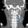
Case #1: A 42 year old female patient was referred by our thoracic surgeon for removal of broken port catheter through an endovascular approach. The patient was diagnosed with bilateral breast cancer 2 years prior. A catheter insertion was performed twenty months prior from the right subclavian vein by our thoracic surgery team to administer chemotherapeutic agents and for blood sampling. Eight months after port placement, it was noted that blood was no longer drawn out of the catheter. To review the catheter, a chest X-ray was performed. This revealed that port catheter was broken from first rib and clavicle region due to POS and that a fractured fragment migrated to the right atrium and right ventricule ( Fig. 1 ). Pinch off Syndrome was suspected given the fracture location. A CT scan was performed to review the port catheter ( Fig. 2 ). After informed consent was obtained, using aseptic measures, a 10 F (French) vascular introducer sheath was placed in the right jugular vein. A 5F snare catheter (AndraSnare®) was introduced from the vascular sheath and after several attempts the proximal end of the fractured port catheter was pulled inside the snare and gradually moved through the sheath ( Fig. 3 ). After removal of the vascular sheath, homestasis was secured by manual compression. No immediate post procedure complication was noted. A control chest x-ray showed no fractured intra-cardiac catheter ( Fig. 4 ).

Related posts:
 A rare and life-threatening case of spontaneous hemopneumothorax presenting with severe hemodynamic instability
 A very rare case of ileocolic and appendiceal intussusception with acute appendicitis
 Hepatic and extra-hepatic hydatid cysts: A case series of radiological and clinical insights
 Mid-term follow-up of a fractured flow diverter in the internal carotid artery
 An atypical multiple autoimmune syndrome: A case report including myocarditis
 Persistence of the hypoglossal artery: A rare case report
Stay updated, free articles. Join our Telegram channel

Full access? Get Clinical Tree


