KEY FACTS
Terminology
- •
Inflammation of epididymis &/or testis
Imaging
- •
Grayscale US
- ○
Epididymis enlarged and hypoechoic
- ○
Testes mildly heterogeneous
- ○
- •
Color Doppler US
- ○
Diffuse or focal hyperemia in body and tail of epididymis ± increased vascularity of testis
- ○
In severe cases can cause vascular compromise and ischemia or infarction
- ○
Reversal of arterial diastolic flow of testis is ominous finding associated with testicular infarction
- ○
- •
Orchitis is usually secondary, occurring in 20-40% of epididymitis due to contiguous spread of infection
Top Differential Diagnoses
- •
Testicular torsion
- •
Testicular lymphoma
- •
Testicular trauma
Pathology
- •
Starts within tail of epididymis → body → testis
Clinical Issues
- •
Commonest cause of acute scrotal pain in adolescent boys and adults (15-35 years)
- •
Males 14-35 years of age: Most commonly caused by Neisseria gonorrhoeae and Chlamydia trachomatis
- •
Scrotal swelling, erythema; fever; dysuria
- ○
Scrotal pain due to epididymitis is usually relieved after elevation of testes (scrotum) over symphysis pubis (Prehn sign)
- ○
Associated lower urinary tract infection and its symptoms, urethral discharge
- ○
- •
Prognosis excellent if treated early with antibiotics; follow-up scans to exclude abscess if no improvement
Scanning Tips
- •
Compare side by side with contralateral asymptomatic side with both color and power Doppler in single image
- ○
Avoid using dual image for side-by-side comparison
- ○
Suggested setting for color Doppler: 5 cm/s
- ○
- •
Head, body, and tail of epididymis should be carefully evaluated
- •
Use high-frequency transducers (9-15 MHz)
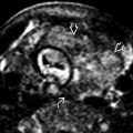
 enveloping the testis posteriorly. Note that the testis
  appears normal in size and configuration.
 compared to the left, and there is a small right hydrocele
  .
.
 .
.
 of the epididymis consistent with acute epididymitis.
 in appearance compared to the left, consistent with right-sided orchitis. In addition, there are associated findings of scrotal wall thickening
  and a small hydrocele
  .
.
 in a patient with a history of epididymo-orchitis. The findings are consistent with a pyocele.


